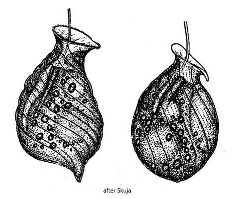
I only very rarely find Urceolus cyclostomus. Mostly the specimens are mixed with detritus flakes. The species is easy to recognize by the funnel-like mouth opening and the distinct spiral striation of the pellicle, which runs counterclockwise. The rod-shaped ingestion organelle becomes visible in squashed specimens (s. fig. 3 a-b). The comparatively large nucleus is located in the rear third of the body (s. fig. 2 d). The flagellum is very long. When swimming, only the distal end rotates. Skuja (1956) also describes specimens with a broadly rounded posterior end (s. drawing above); in my population all specimens had a pointed posterior end (s. fig. 2 b).
Fig. 1 a-d:Urceolus cyclostomus. L = 35 µm. Different focal planes of a freely swimming specimen. CV = contractile vacuole, F = flagellum. Obj. 100 X.
Fig. 2 a-d:Urceolus cyclostomus. L = 37 µm. Different focal planes of a second freely swimming specimen. CV = contractile vacuole, F = flagellum, IO = rod-shaped ingestion organell, Nu = nucleus, RE = reservoir. Obj. 100 X.
Fig. 3 a-b:Urceolus cyclostomus. The ingestion organell (IO) in a squashed specimen. Nu = nucleus, RE = reservoir. Obj. 100 X.