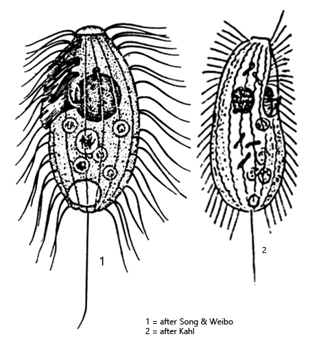
dorsal side convex, oral apparatus slightly indented
obliquely truncated frontal plate
oral apparatus with 3 membranelles in anterior third
undulating membrane on right side
macronucleus spherical, one spherical micronucleus
contractile vacuole terminal
extrusomes rod-shaped, inconspicuous
cytoplasm often darkly spotted due to crystalline inclusions
10–14 longitudinal rows of cilia
paired cilia in anterior third
one caudal cilium, half body length
Uronema nigricans
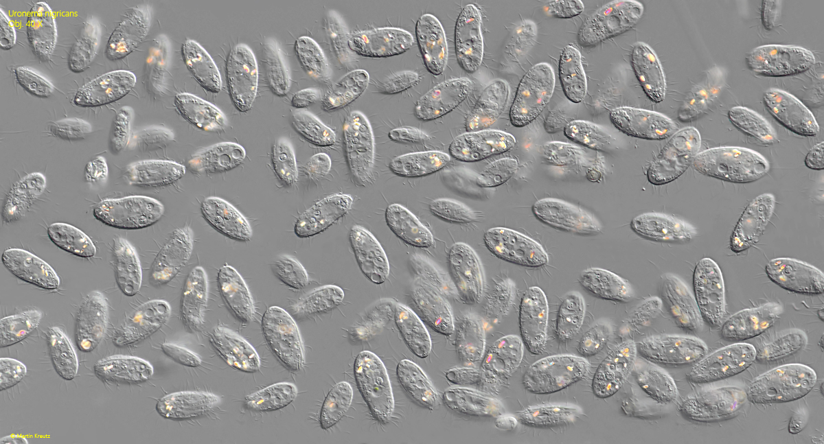
I find Uronema nigricans rarely in the Simmelried and very frequently in the pond at the waste disposal company Constance. In particular, cutting the stems of the fringed water lily (Nymphoides peltata) growing there into pieces and leaving them to decay in a Petri dish results in strong mass developments of Uronema nigricans.
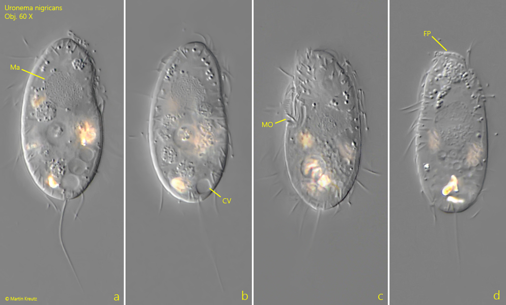
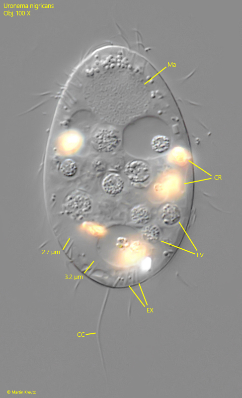
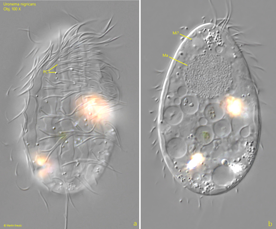
At medium magnification, one can see a characteristic indentation in the anterior third of the body (s. fig. 3 a-c), where the mouth apparatus is located. One can also clearly see a frontal plate (s. fig. 3 d), the slightly subterminal contractile vacuole (s. fig. 3 b), and a caudal cilium (s. fig. 5), which is often angled. Only at high magnification can one see that the cilia in the anterior third are arranged in pairs and are also more densely spaced here than in the posterior part of the body (s. fig. 6 a). The round macronucleus is located in the anterior third (s. figs. 5 and 6 b). The micronucleus is difficult to see (s. fig. 6 b). Numerous highly refractive crystals are often present in the cytoplasm, which can make examining the specimens difficult (s. fig. 5).
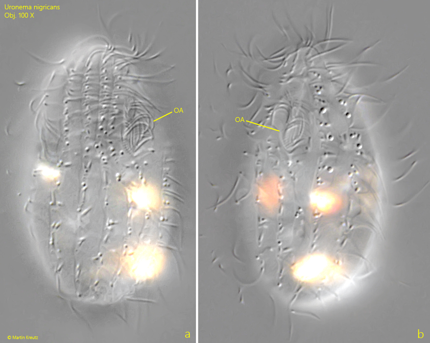
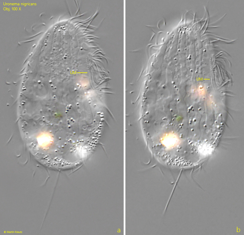
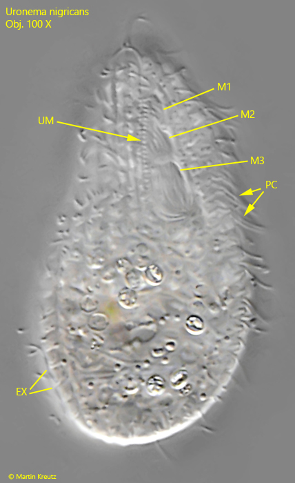
The structure of the oral apparatus is difficult to discern without silver impregnation, as it is also very small (s. fig. 7 a-b). An undulating membrane is located on the right side, and three adoral membranelles lie above the actual mouth opening (s. figs. 8 a-b and 9).
Fig. 1:Uronema nigricans. A mass development in a sample with decaying water lilly stems. Obj. 40 X.
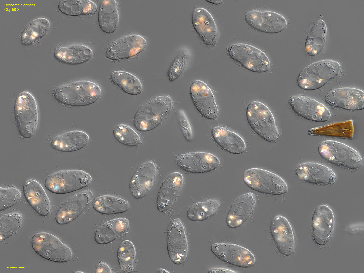
Fig. 2:Uronema nigricans. L = 31–42 µm. Slightly squashed specimens in the same sample as shown in fig. 1. Obj. 60 X.
Fig. 3 a-d:Uronema nigricans. L = 40–42 µm. A slightly specimen from right (a, b) and a second specimen from left und from dorsal (c, d). CV = subterminal contractile vacuole, FP = frontal plate, Ma = macronucleus, MO = mouth opening. Obj. 60 X.
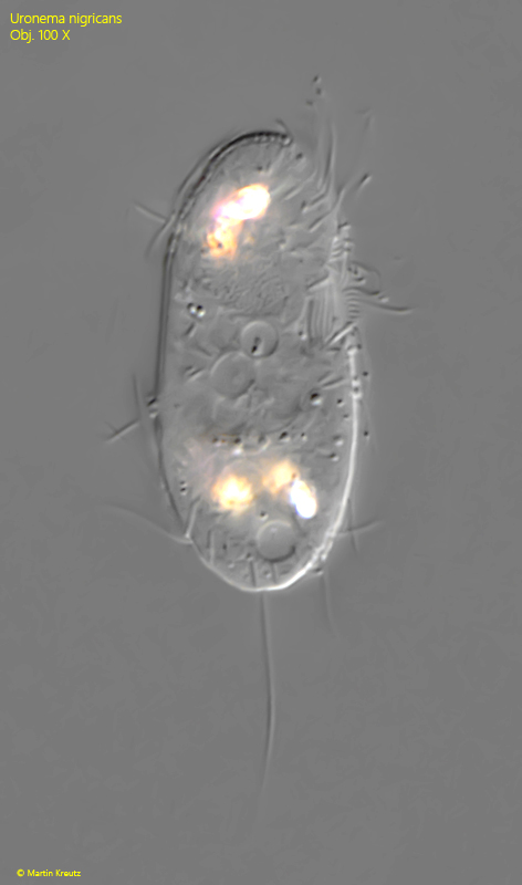
Fig. 4:Uronema nigricans. L = 40 µm. A freely swimming specimen from right. Obj. 100 X.
Fig. 5:Uronema nigricans. A squashed specimen. The rod-shaped extrusomes (EX) are 2.7–3.2 µm long. CC = caudal cilium, CR = refractive crystals, FV = food vacuoles, Ma = macronucleus. Obj. 100 X.
Fig. 6 a-b:Uronema nigricans. Two focal planes of a squashed specimen. Note the paired cilia (PC) in the anterior third and the spherical macronucleus (Ma). Mi? = probably the micronucleus. Obj. 100 X.
Fig. 7 a-b:Uronema nigricans. A second squashed specimen in ventral view with focal plane on the oral apparatus (OA). Obj. 100 X.
Fig. 8 a-b:Uronema nigricans. The same specimen as shwon in fig. 7 a-b with focal plane on the unulating membrane (UM) on the right side of the oral apparatus. Obj. 100 X.
Fig. 9:Uronema nigricans. A well-fed specimen from ventral. The threee adoral membranelles (M1–M3) are visible and the undulating membrane (UM) of the right side. EX = extrusomes, PC = paired cilia. Obj. 100 X.