Urostyla grandis Ehrenberg, 1830

Most likely ID: n.a.


Synonym: n.a.


Sampling location: Pond of the wase disposal company Constance


Phylogenetic tree: Urostyla grandis


Diagnosis: 

  • body elliptical, almost parallel-sided, dorso-ventrally flatttened
  • length 250–400 µm, width 80–120 µm
  • adoral zone 35–40 % of body length
  • one endoral and one paroral membrane
  • about 70–150 macronuclear pieces scattered in cytoplasm
  • about 40 spherical micronuclei
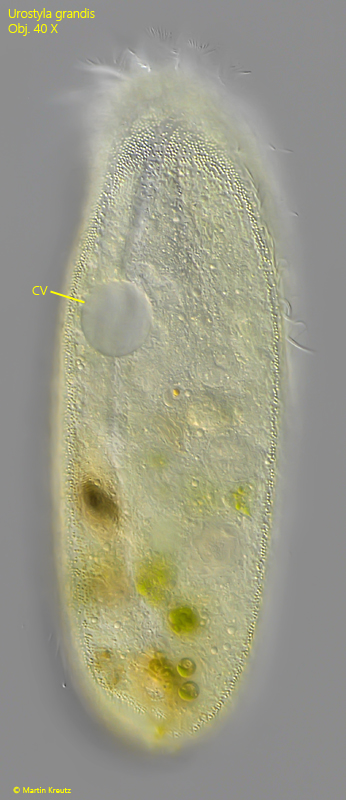
  • one contractile vacuole, in anterior third, left margin
  • ventral several rows of yellowish granules (sometimes granules shaped elliptical)
  • ventral 10–12 longitudinal rows of cirri (between granules)
  • 10–15 transverse cirri on oblique row
  • anterior 18–27 obliquely arranged pairs of cirri
  • cilia free zone below mouth opening
  • dorsal 3–4 rows of short cirri
  • caudal cirri absent
Urostyla-grandis
Urostyla grandis

Urostyla grandis is a very conspicuous ciliate due to its size of about 300 µm, which I find only occasionally. However, I was able to study a very large population in a prepared sample from the pond of waste disposal company Constance. To do so, I cut stems of the yellow water lily (Nuphar lutea) from this pond into pieces and placed them in a Petri dish with water from the collection site. The population of Urostyla grandis appeared in this sample after about 1 week. All the images shown below are of specimens from this sample.


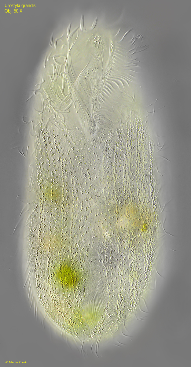
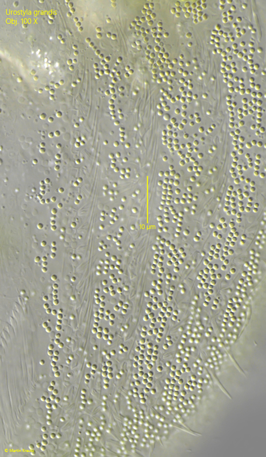
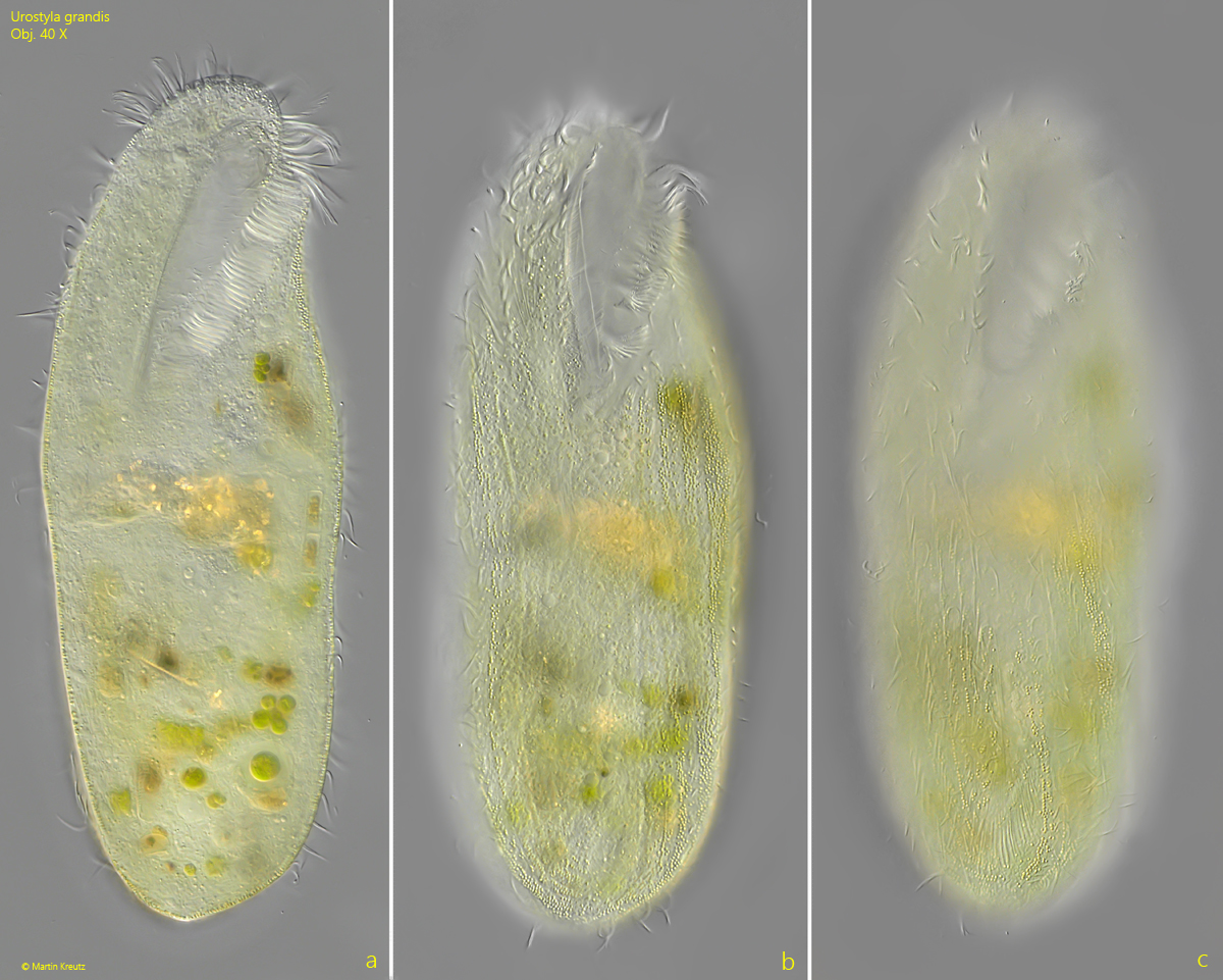
Urostyla grandis is a hypotrich ciliate with a yellow coloration. In my population, the coloration was lemon yellow (s. fig. 1 a-c). However, it can also be greenish or brownish. The coloration is caused by cortical granules, which are arranged in longitudinal stripes (s. figs. 2 and 3). The granules are about 1 µm in size. In my population, the granules were spherical in shape, but they can also take on elliptical forms.


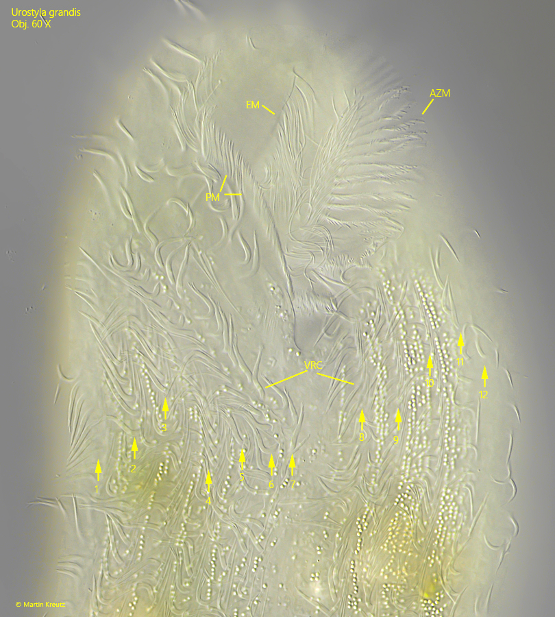
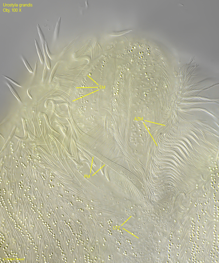
Between the bands of granules, rows of cirri run along the ventral side. I was able to identify about 12 by light microscopy (s. fig. 5). The ciliature around the oral apparatus is complicated and difficult to interpret without silver impregnation. The oral apparatus consists of an adoral zone on the right side and an endoral and a paroral membrane on the left side (s. figs. 5 and 6). Below the oral apparatus is a zone that is free of cirri (s. fig. 6).


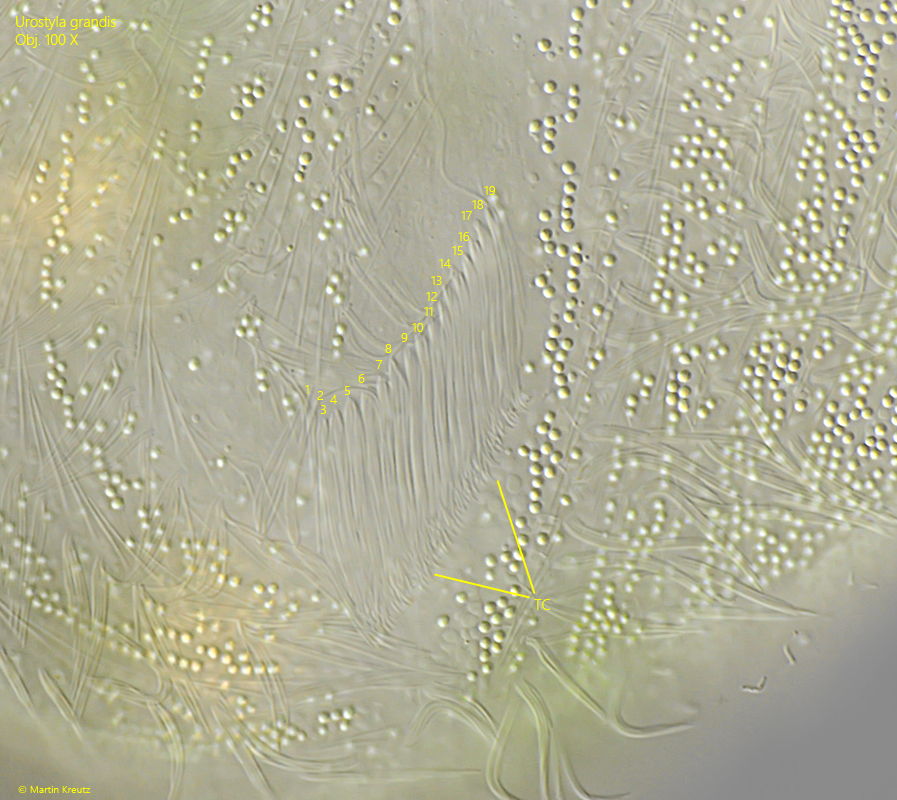
In the posterior quarter, there is an oblique row of transverse cirri on the ventral side. According to Foissner, Blatterer, Berger and Kohmann (1991), there should be 10–15 cirri. There were 19 in my population (s. fig. 6).


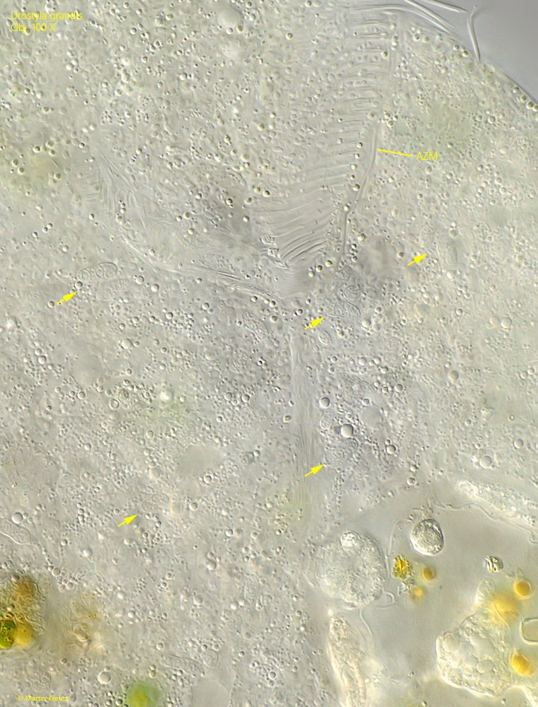
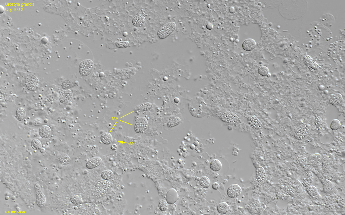
An important diagnostic feature of Urostyla grandis is its nuclear apparatus. In Urostyla grandis, the macronucleus consists of up to 150 small pieces distributed throughout the cytoplasm, each only 5–10 µm in size (s. figs. 8 and 9). These pieces are easy to overlook in freely swimming or only slightly compressed specimens. They can be identified precisely only in heavily squashed specimens (s. fig. 9).


The similar species Paraurostyla weissei has two separate macronuclei, each with an attached micronucleus. This makes Urostyla grandis and Paraurostyla weissei easy to distinguish from one another. The other similar species, Pseudourostyla cristata, has extrusomes, which are absent in Urostyla grandis.

Urostyla-grandis

Fig. 1 a-c: Urostyla grandis. L = 372 µm. Three focal planes of a freely swimming and intensely yellow-colored specimen. Obj. 40 X.

Urostyla-grandis

Fig. 2: Urostyla grandis. L = 336 µm. The ventral side of a slightly squashed specimen with the rows of yellow-colored granules. Obj. 60 X.

Urostyla-grandis

Fig. 3: Urostyla grandis. The yellow cortical granules in detail. They have a diameter of about 1 µm. Obj. 100 X.

Urostyla-grandis

Fig. 4: Urostyla grandis. L = 323 µm. A specimen from dorsal with the contractile vacuole (CV) on the left side in the anterior third. Obj. 40 X.

Urostyla-grandis

Fig. 5: Urostyla grandis. The oral apparatus possesses an adoral zone of membranelles (AZM) on the right side as well as an endoral (EM) and paroral membrane (PM) on the left side. The ventral rows of cirri (VRC, 1–12) run between the rows of cortical granules. Obj. 60 X.

Urostyla-grandis

Fig. 6: Urostyla grandis. The oral apparatus of a squashed specimen. Note the cirri free zone (CFZ) below the oral apparatus. AZM = adoral zone of membranelles, EM = endoral membrane, PM = paroral membrane. Obj. 100 X.

Urostyla-grandis

Fig. 7: Urostyla grandis. The transverse cirri (TC) are arranged in an oblique row. In this specimen 19 transverse cirri are present (1–19). Obj. 100 X.

Urostyla-grandis

Fig. 8: Urostyla grandis. In the cytoplasm numerous macronuclear pieces are scattered (arrows). Because of their small size of 5–10 µm, they are easy to overlook. Obj. 100 X. 

Urostyla-grandis

Fig. 9: Urostyla grandis. The macronuclear pieces (Ma) in a squashed specimen. Mi? = probably a micronucleus. Obj. 100 X.