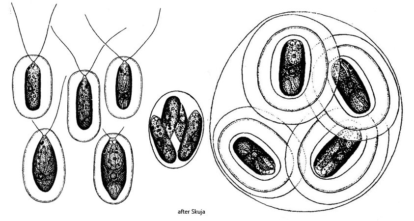
two polar flagella, three-quarters to four-fifths of body length
chloroplast parietal, sac-shaped
one central pyrenoid
nucleus in anterior third
two anterior contractile vacuoles
one eyespot in anterior third
form resting stages with distinctly layered sheath
Vitreochlamys cylindrica
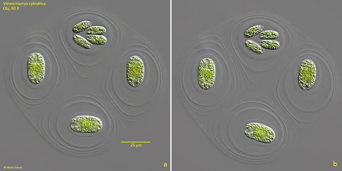
So far, I have only found Vitreochlamys cylindrica in the pond of the convent Hegne. However, I did not find the flagellated stages, which have two flagella and are surrounded by a gelatinous envelope, but rather the resting stages, which are particularly characteristic of Vitreochlamys cylindrica. In the resting stages, 4–16 cells lie together in a clearly layered gelatinous envelope (s. fig. 1 a-b). The cells are not flagellated but immobile. Cell division also takes place in these resting stages, from which 4 to 8 daughter cells emerge.
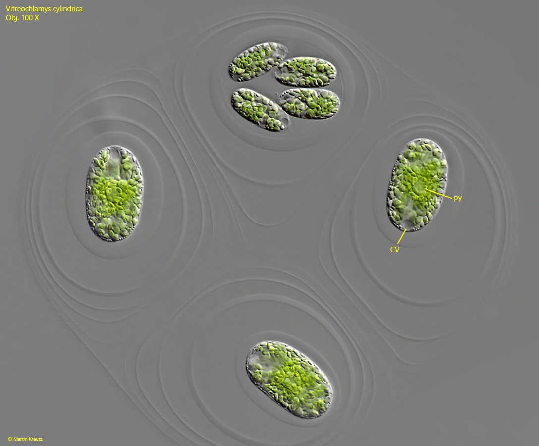
In the resting stages of Vitreochlamys cylindrica that I found, all cells were filled with starch grains. As a result, I could neither see the eyespot nor the position of the nucleus. Of the two apically located contractile vacuoles, only one was always visible because they pulsate alternately. The adult cells were 25–27 µm long, which corresponds well to the data from Skuja (1948).
Fig. 1 a-b:Vitreochlamys cylindrica. L = 27 µm (of adult cells). Two slightly different focal planes of a resting stage with 3 adult cells and the formation of 4 daughter cells. Note the distinct layers of the gelationous sheath covering the cells. Obj. 60 X.
Fig. 2:Vitreochlamys cylindrica. L = 27 µm (of adult cells). The same resting stage as shown in fig. 1 a-b at higher magnification. CV = contractile vacuole, PY = pyrenoid. Obj. 100 X.