contracted cells almost spherical with distinct anterior bumps
length 50–160 µm, width 35–100 µm
cells appears dark due to refractive oil droplets in the cytoplasm
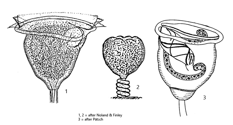
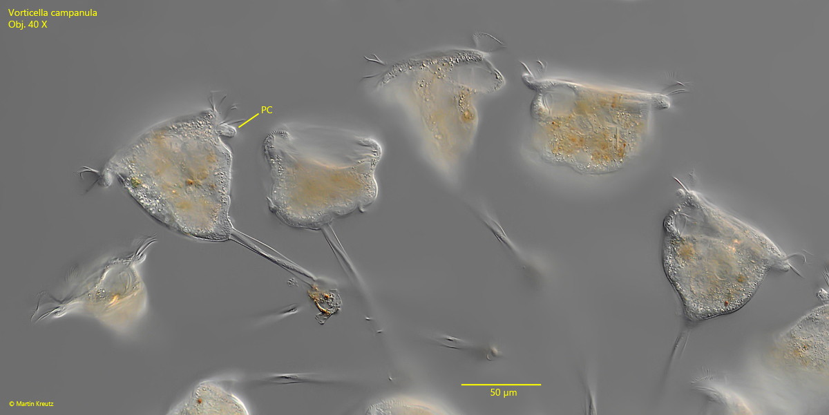
peristome protruding clearly body margin
macronucleus J-shaped in longitudinal axis
one contractile vacuole near ventral wall of oral funnel
pellicle finely striated transversely, about 72 lines
stalk contracts in tightly helical line
solitary or in pseudocolonies
Vorticella campanula
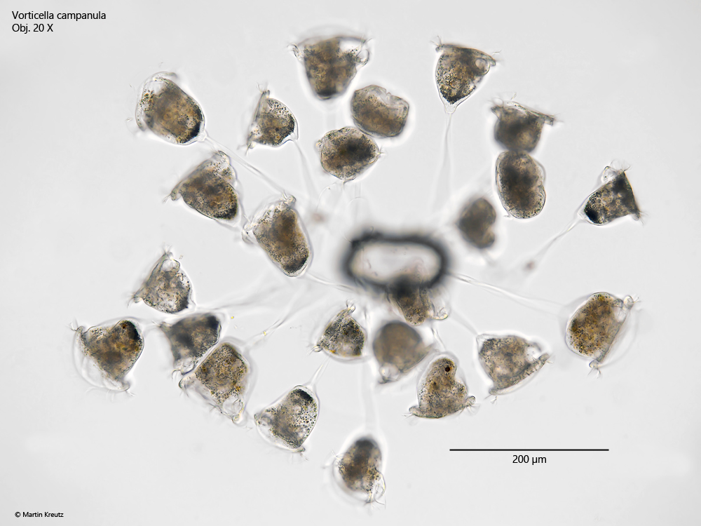
Vorticella campanula is one of the most common members of the genus and I find the species in many of my sampling sites. Vorticella campanula is easy to recognize even at low magnification because the cells are very large at about 100 µm in length and because they appear dark, sometimes even black, especially in bright field illumination (s. figs. 1 and 2). This is due to the large quantities of oil droplets that fill the entire cyptoplasm (s. fig. 6). At the posterior end of the cell, at the base of the stalk, the concentration of oil droplets is particularly high. The cells are bell-shaped and about as wide as they are long. The peristomal collar clearly protrudes beyond the margin of the body (s. fig. 3). There is only one contractile vacuole, which can usually only be clearly recognized in squashed specimens (s. fig. 6). It is located in the anterior third, attached to the oral funnel, into which it also empties. The striation of the pellicle is fine and can only be recognized at high magnification (s. fig. 5).
Fig. 1:Vorticella campanula. L = 72–86 µm. A pseudocolony of 20–30 specimens in brightfield illumination. Note the dark color of the cells due to refractive oil droplets in the cytoplasm. Obj. 20 X.
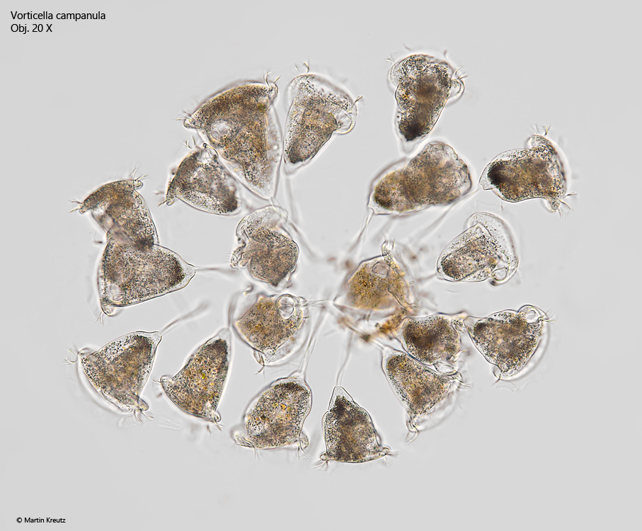
Fig. 2:Vorticella campanula. L = 91–122 µm. A second, smaller pseudocolony in brightfield illumination. Obj. 20 X.
Fig. 3:Vorticella campanula. L = 75–91 µm. Some extended specimens of a pseudocolony. PC = peristomal collar. Obj. 40 X.
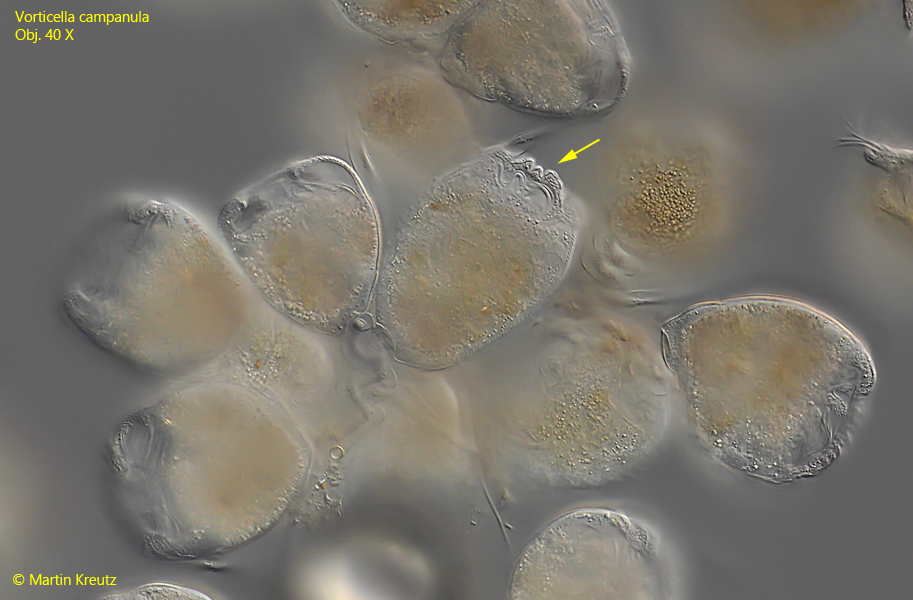
Fig. 4:Vorticella campanula. In contracted cells the lip of the peristome is folded to distinct bumps (arrow). Obj. 40 X.
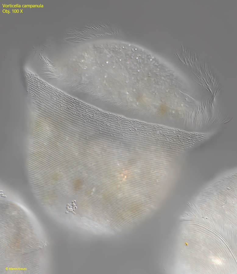
Fig. 5:Vorticella campanula. The fine, transverse striation of the pellicle. L = 91–122 µm. A second, smaller pseudocolony in brightfield illumination. Obj. 100 X.
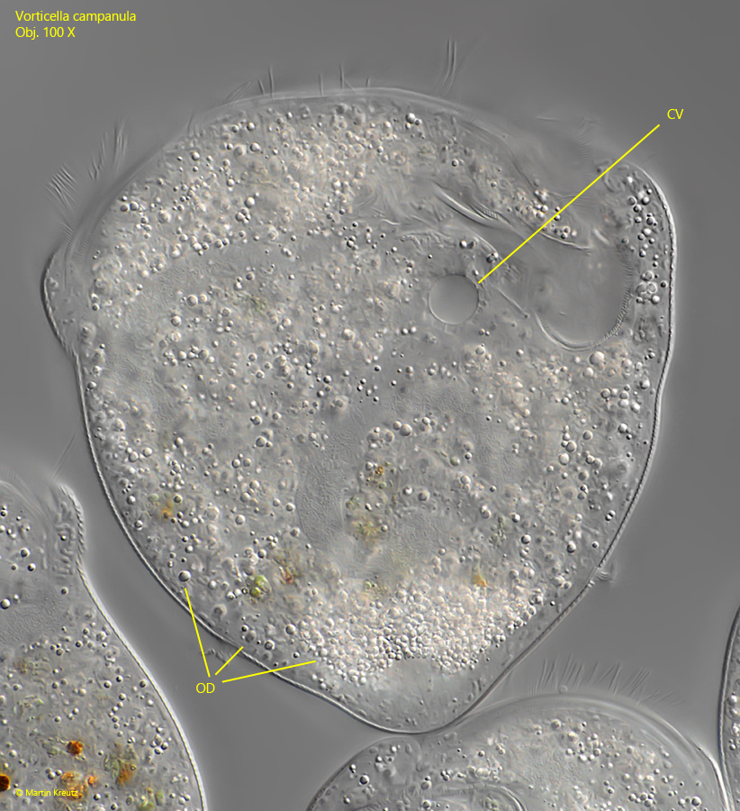
Fig. 6:Vorticella campanula. In this squashed specimen the single contractile vacuole (CV) is visible. The cytoplasm is filled with refractive oil droplets (OD). Obj. 100 X.
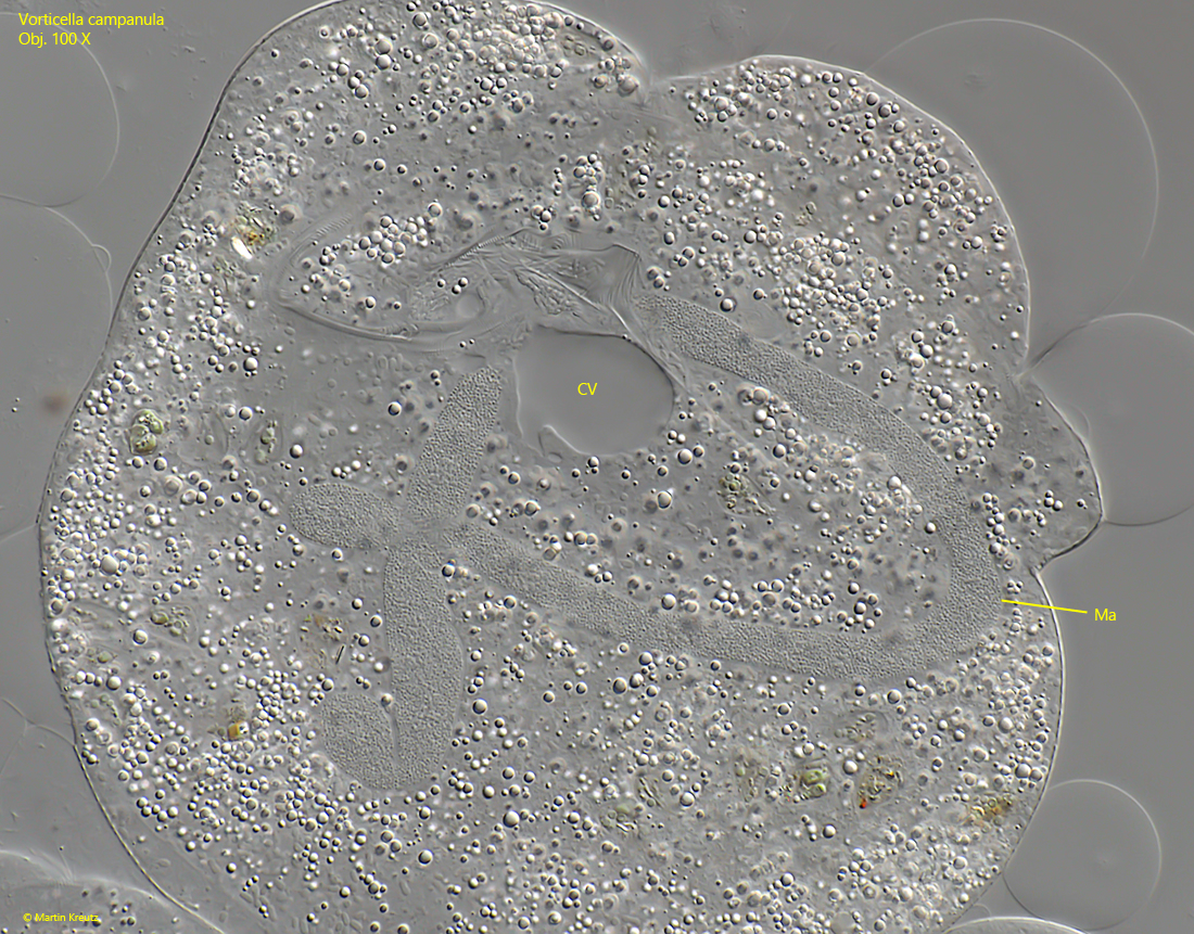
Fig. 7:Vorticella campanula. The J-shaped macronucleus (Ma) in a strongly squashed specimen. CV = contractile vacuole). Obj. 100 X.
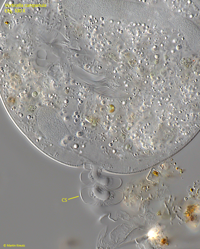
Fig. 8:Vorticella campanula. The tightly helical stalk (CS) in a contracted specimen. Obj. 100 X.