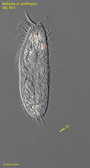
So far, I have only found 3 specimens of Wallackia cf. schiffmanni. The first two specimens were found in September 2008 in Simmelried. The third specimen was found only 14 years later, in July 2022, also in Simmelried.
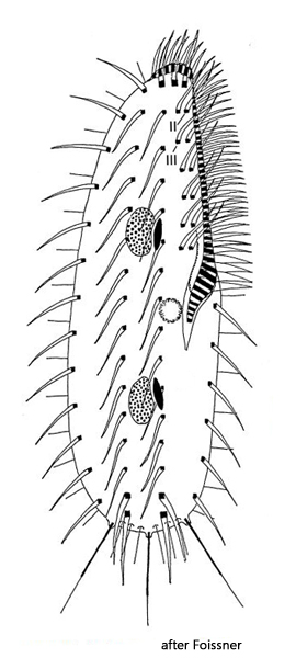
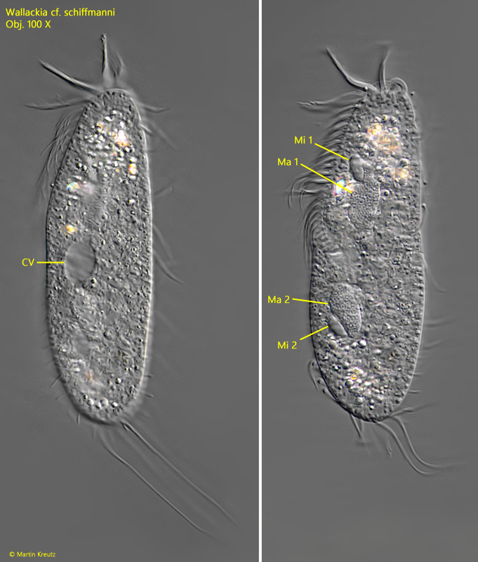
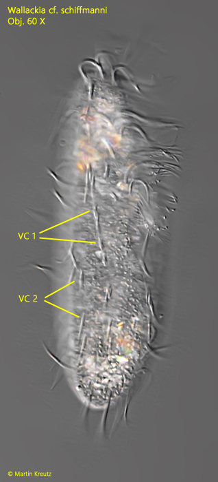
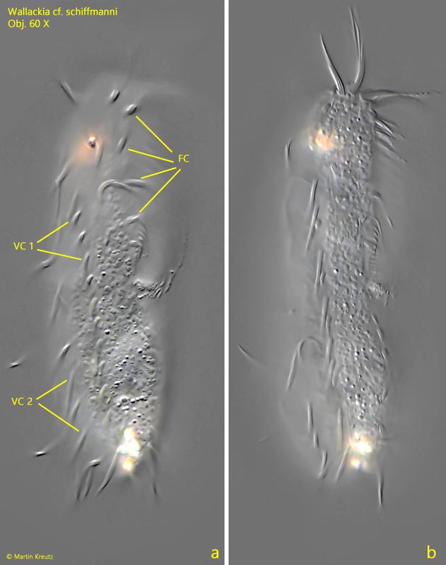
Within the genus Wallackia, only three species have been recorded so far. These hypotrich ciliates share the characteristic that they all have elongated caudal cirri and two parallel longitudinal rows of ventral cirri. The adoral zone extends to the middle of the body in all three species, and the contractile vacuole is located on the right side, emptying directly into the cytopharynx. There are always two macronuclei and two micronuclei present.
The species Wallackia elegans has a distinctly tapered posterior end and is only about 70 µm long. I therefore excluded this species, as my specimens were between 84–110 µm long and had a broadly rounded posterior end. This leaves the two similar species Wallackia schiffmanni and Wallackia bujoreani. The latter species is said to live terrestrially and also only reaches 70 µm in length. Thus, Wallackia schiffmanni remains, for which a length of 85–100 µm is given (Foissner, 1976) and which also occurs limnetically.
However, my findings differ from Wallackia schiffmanni particularly in the length and arrangement of the caudal cirri compared to Foissner’s original description. According to that, there should be only 3 caudal cirri, which are widely spaced and arise from short, tubular extensions, as Foissner drew them (s. drawing above). A specimen of Wallackia schiffmanni found by Bruce Taylor (s. link below) clearly shows the three spread caudal cirri. In contrast, the specimens of my population had at least 4 caudal cirri, which were between 25–52 µm long and soft and flexible. They also did not arise from tubular extensions. Additionally, Wallackia schiffmanni is supposed to have a fringe of about 3 µm long, rod-shaped proto-extrusomes, which I could not detect in my specimens either.
As an alternative, Wallackia bujoreani remains, which Berger (2011) as well as Hemberger (1982) believe might be synonymous with Wallackia schiffmanni and possibly not only terrestrial. In Wallackia bujoreani, the caudal cirri are indeed closer together, but there are said to be only three.
Due to the length of the specimens in my population and the limnic habitat, I therefore consider Wallackia schiffmanni most likely; however, because of the deviations from the original descriptions described above, I would like to designate the species as Wallackia cf. schiffmanni.
More images and information on Wallackia schiffmanni: Bruce Taylor-iNaturalist-Wallackia schiffmanni