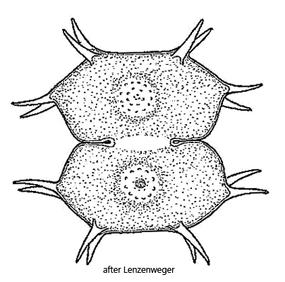
semi-cells transverse elliptical or elongated hexagonal
slightly convex or straight sides and apex
each semi-cells with 4 pairs of simple spines
length 40–75 µm (without spines)
center of semi-cells with variable ornamentation of warts
one parietal chloroplast with two pyrenoids per semi-cell
Xanthidium antilopaeum var. ornatum
So far I have only found Xanthidium antilopaeum var. ornatum in the Paradieswiesen (Austria) and in the Simmelried. In the Simmelried, however, the species occurs only very rarely.
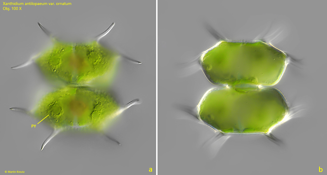
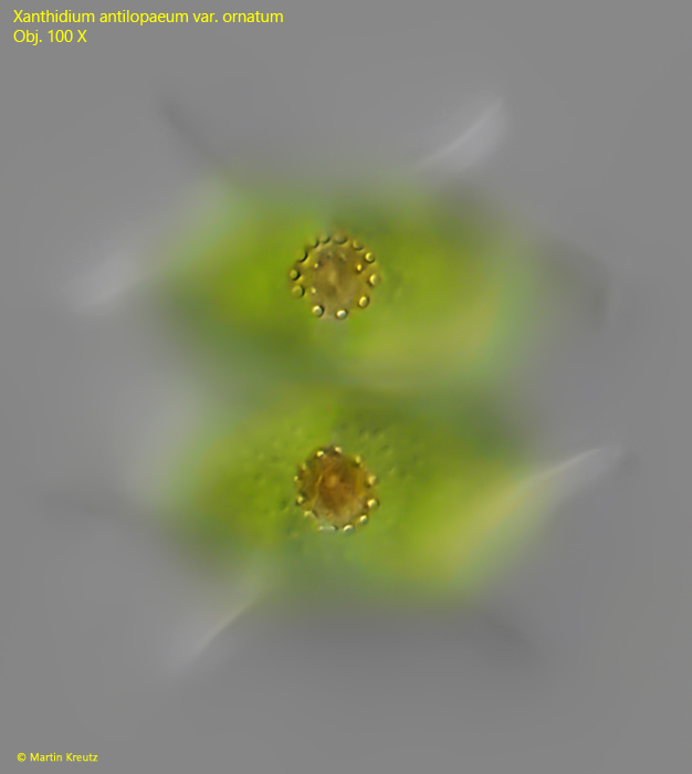
Each semi-cell of Xanthidium antilopaeum has 4 pairs of straight or slightly curved spines. The equatorial spines are usually bent towards the center of the cell (s. fig. 1 a). In the center of each half cell there is an ornamentation of round or elongated warts, which can be very variable. In the specimens of my population it was a ring of round tubercles on a brownish colored field (s. fig. 2).
Xanthidium antilopaeum var. ornatum can easily be distinguished from the similar species Xanthidium cristatum and Xanthidium fasciculatum by the number of spines per semi-cell. While Xanthidium antilopaeum var. ornatum has 8 spines per half cell (4 pairs), the other two species have 10 spines (4 pairs and 2 solitary spines each).
Fig. 1 a-b:Xanthidium antilopaeum var. ornatum. L = 57 µm (without spines). Two focal planes of a specimen found in the Paradieswiesen. PY = pyrenoid. Obj. 100 X.
Fig. 2:Xanthidium antilopaeum var. ornatum. L = 57 µm (without spines). Focal plane on the ornamentation of ring-shaped arranged warts in the center of the semi-cells. Obj. 100 X.