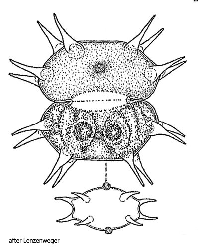
semi-cells transverse elliptical or elongated hexagonal
slightly convex or straight sides and apex
each semi-cells with 4 pairs of straight spines
length 43–55 µm (without spines)
center of semi-cells with spherical wart
both sides of isthmus with rows of small warts
one parietal chloroplast with two pyrenoids per semi-cell
Xanthidium basiornatum
So far, I have found only one specimen of Xanthidium basiornatum in July 2026, in a 6-week-old sample from the Schwemm Moor in Austria.
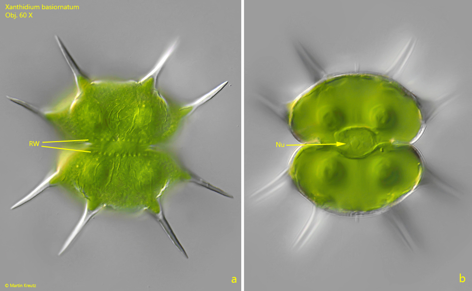
Xanthidium basiornatum closely resembles Xanthidium antilopaeum, but the 8 spines per semi-cell are all straight and long. In addition, they arise from the semi-cells almost vertically. Furthermore, Xanthidium basiornatum has a linear row of small warts above and below the isthmus (s. fig. 1 a). According to Lenzenweger (1997), there should also be a spherical wart in the middle of each semi-cell, which I was unable to detect in my specimen.
More images and information on Xanthidium basiornatum:
Fig. 1 a-b:Xanthidium basiornatum. L = 55 µm (without spines). Two focal planes of the specimen found in July 2026 in the Schwemm. Note the rows of warts (RA) above and below the isthmus and the central nucleus (Nu). Obj. 60 X.
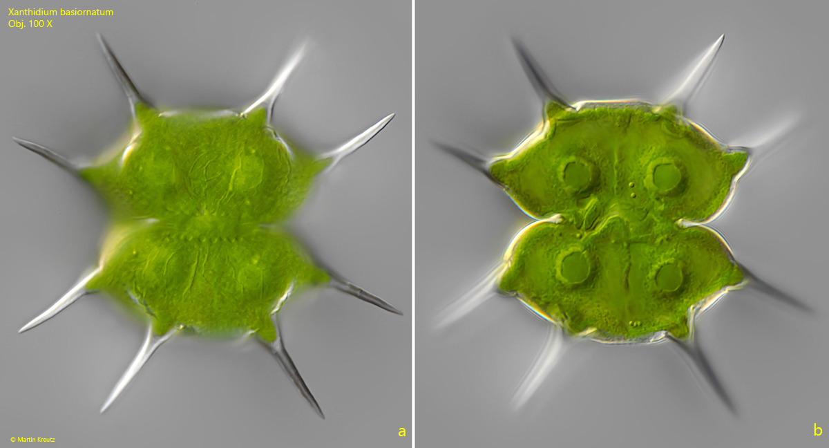
Fig. 2 a-b:Xanthidium basiornatum. L = 55 µm (without spines). The same specimen as shown in fig. 1 a-b at higher magnification. Obj. 100 X.