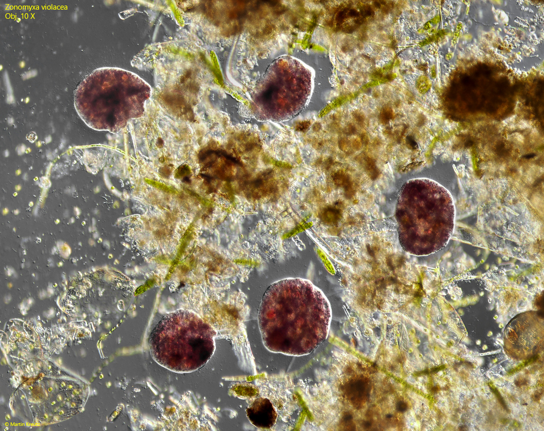
So far, I have only found Zonomyxa violacea once in July 2017 in the Sima Moor in Austria. Especially in older samples with a thin layer of detritus and algae at the bottom of the containers, the specimens multiplied well. The population remained constant for several weeks.
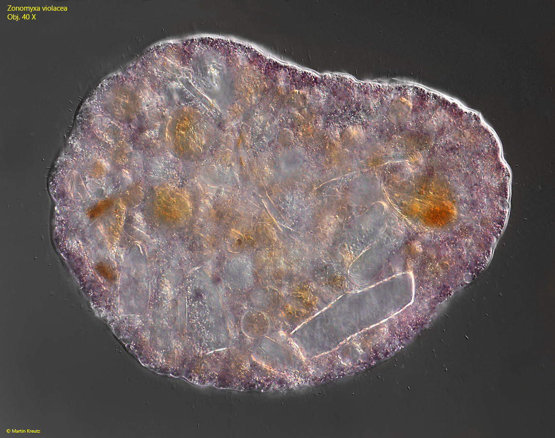
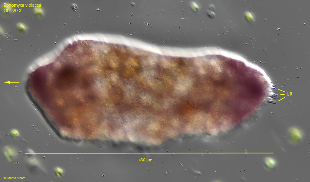
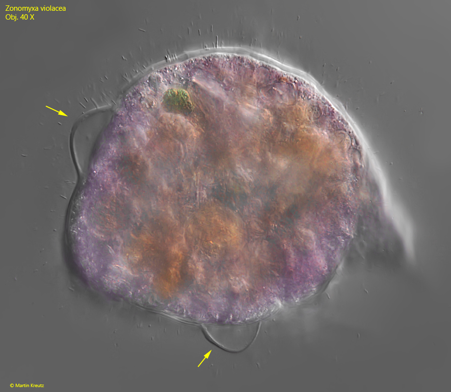
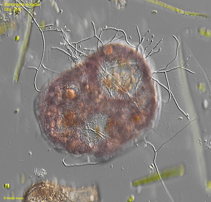
The specimens of Zonomyxa violacea moved very little and then only very slowly. Most of the time, the body was rounded or knobby. The rounded specimens had a diameter of 140–220 µm. Only very rarely did I find specimens in a limax form (s. fig. 6), which were then over 400 µm long. The specimens phagocytized large amounts of detritus mixed with algae (s. figs. 1, 2 and 3). Everything was phagocytized completely unspecifically, which reminded me of the feeding behavior of Pelomyxa palustris. Through this feeding behavior, the specimens burrowed deeper and deeper into the detritus.
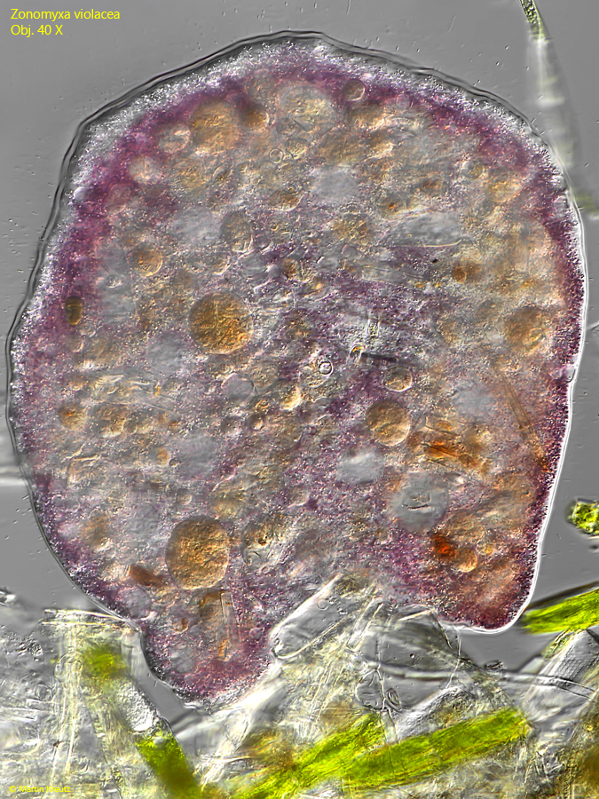
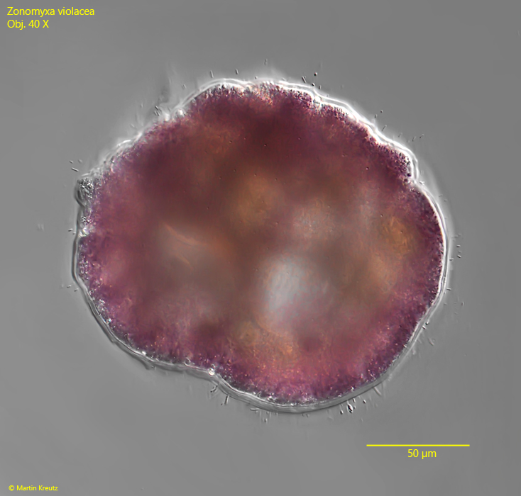
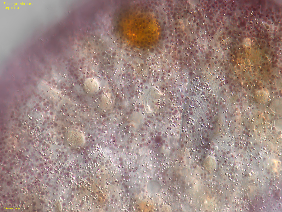
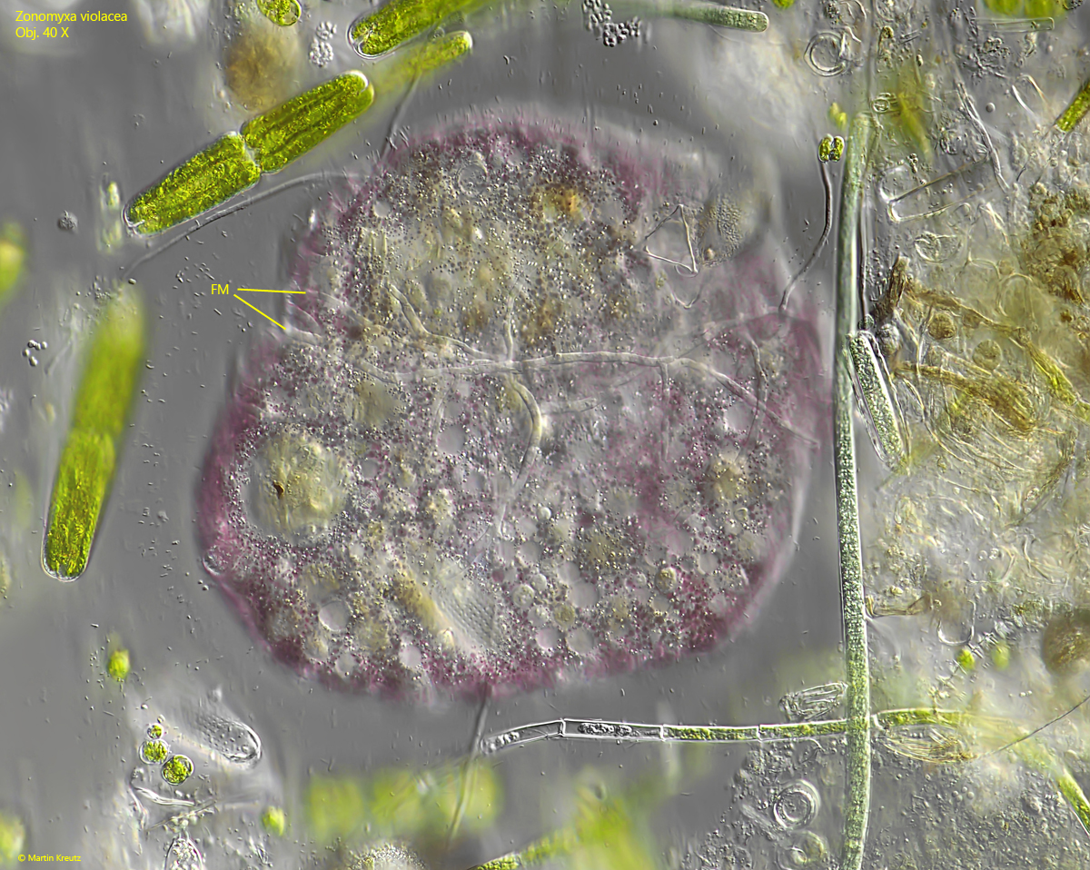
Although the cytoplasm was always filled with phagocytosed detritus and algae, all specimens showed a distinct violet coloration, which sometimes tended slightly towards reddish. This coloration is caused by large amounts of violet-colored vesicles with a diameter of 1-5 µm, which are distributed throughout the cytoplasm but often concentrate near the cell surface (s. fig. 7). In burst specimens, I observed that the color of the vesicles quickly change to brown or yellow-brown when they leak out. Possibly, the responsible pigment is sensitive to pH changes or oxygen.
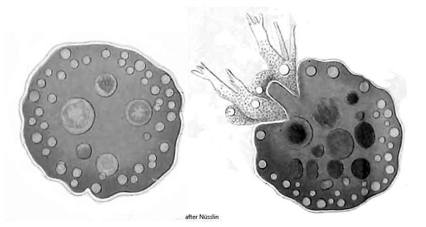
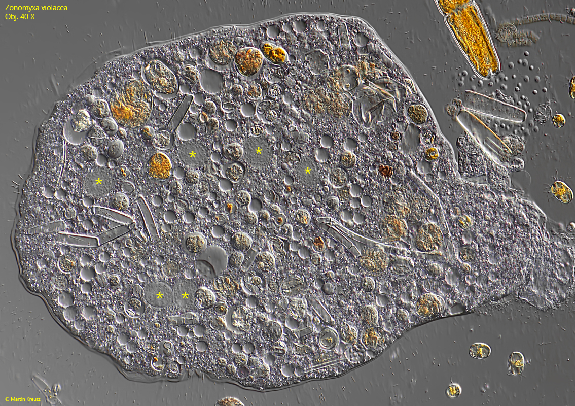
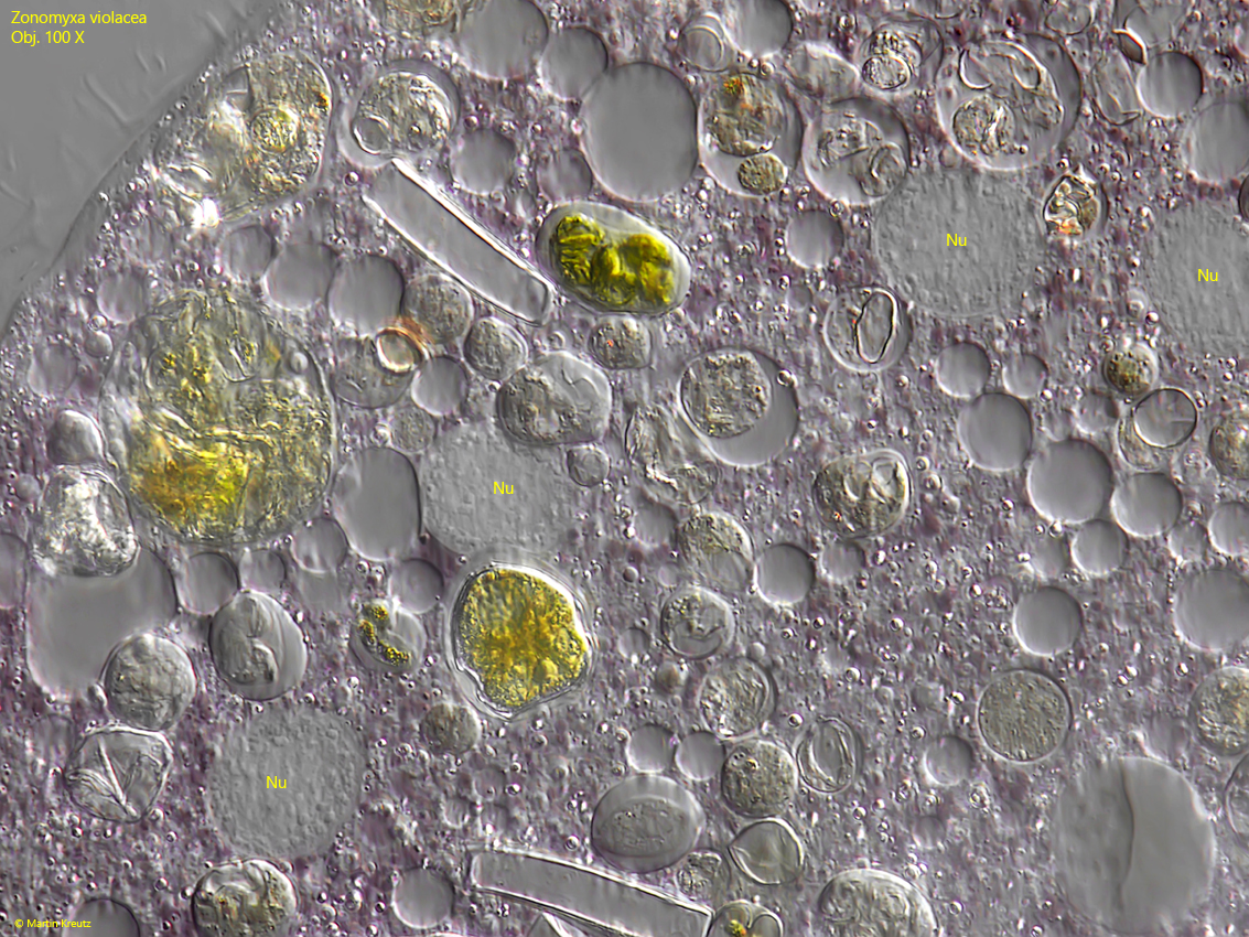
The nuclei of Zonomyxa violacea can only be recognized in strongly squashed specimens due to the large amounts of detritus in the body. In my population, there were always 6–10 nuclei, which had a diameter of 18–22 µm (s. figs. 8 and 9). In one case, I also found a specimen with nuclei measuring 35 µm. These dimensions correspond to the observations of Penard (1906), who reported 20 µm, and Siemensma (s. link below), who measured 28 µm. Opitz (2017) found 6 nuclei per specimen in a population from the Hochmoor Schweizer Alm in Austria, and Siemensma (see link below) found an average of 7 nuclei. Specimens with 32 nuclei, as described by Penard (1906), have not been found again since.
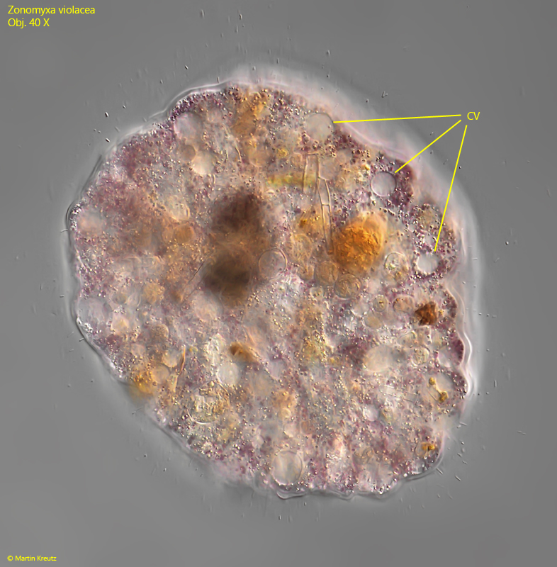
The numerous contractile vacuoles in Zonomyxa violacea are located near the cell surface. They can be easily seen there (s. fig. 10). There are about 20–60 contractile vacuoles per cell.
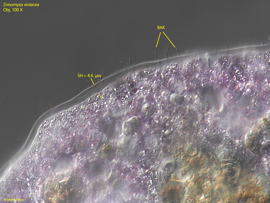
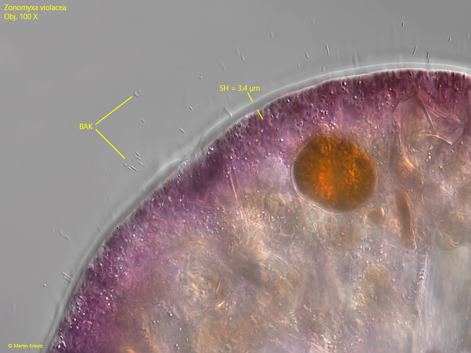
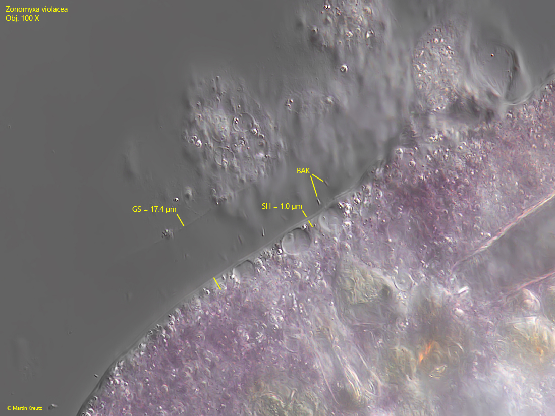
The colorless shell of Zonomyxa violacea consists of a soft and flexible, chitinous layer. In some specimens, I was able to discern layering (s. fig. 11). In some cases, I also observed how the shell clearly detached from the cell body, creating blister-like elevations (s. fig. 14). This detachment of the shell may be caused by the movement of the cell body, which the shell cannot adapt to. However, this phenomenon illustrates that the shell lies loosely against the plasma body. The thickness of the shell in Zonomyxa violacea appears to be subject to some variability. I measured a range of 1–5 µm (s. figs. 11, 12 and 13).
Above this chitinous shell, bacteria were visible in all specimens, arranged radially to the amoeba body. The matrix in which these bacteria lie was in the most cases not visible in DIC. In my opinion, the presence of these bacteria is caused by a gelatinous sheath cover the amoeba body, as is actually described for Amphizonella violacea. In one case I coud recognize the margin and dimension of this gelatinous sheath (s. fig. 13). From this observation and from the position and arrangement of the bacteria, one can conclude that this gelatinous sheath in Zonomyxa violacea has a thickness of about 10–40 µm. How this gelatinous layer is formed remains unclear. It may possibly be a mucilaginous coating of the shell, similar to how the cell walls of algae can become mucilaginous.
The intense violet coloration of Zonomyxa violacea is also found in the very similar species Amphizonella violacea. It is difficult to distinguish these species. Meisterfeld and Badewitz (2006) provide a redescription of Amphizonella violacea, in which the authors explain the main differences from Zonomyxa violacea:
- Amphizonella violacea has only one nucleus, while Zonomyxa violacea has 4–32
- a gelatinous sheath over the shell is absent in Zonomyxa violacea
- Zonomyxa violacea has a thinner shell than Amphizonella violacea
- the habitat of Zonomyxa violacea is Sphagnum bogs, while Amphizonella violacea lives in xerophilous mosses (on roofs, walls, or trees) with strongly fluctuating water content
However, in my opinion, some of these characteristics are very unspecific. The habitats overlap, as Amphizonella violacea as well as Zonomyxa violacea can be found in Sphagnum bogs. The thickness of the shell is very variable in both species, and here too the measurements overlap. Finally, the gelatinous sheath can also be absent in Amphizonella violacea. The number of nuclei remains the most reliable characteristic. Zonomyxa violacea has constantly >=4 nuclei, while Amphizonella violacea always seems to be uninucleate.
More images and information on Zonomyxa violacea: Ferry Siemensma-Microworld-Zonomyxa violacea