cells in the colonies are separated from each other
no visible gelatinous sheath
only few visible division stages in the colonies
granules in the cells are scattered, with some distinct dark granules per cell
No drawings from previous authors available.
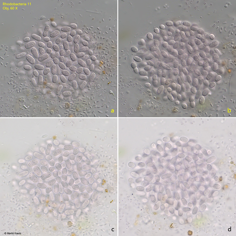
Fig. 1:Rhodobacteria 11. L = 5–10 µm. A slightly squashed colony in DIC (a, b) and in brightfield illumination (c, d). All cells are separated from each other. No gelatinous sheath is visible. Obj. 60 X.
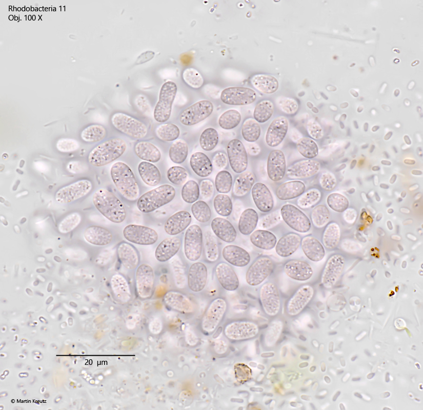
Fig. 2:Rhodobacteria 11. L = 5–10 µm. The same colony as shown in fig. 1. Obj. 100 X.
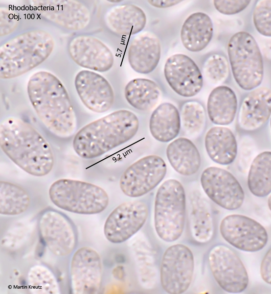
Fig. 3:Rhodobacteria 11. L = 5–10 µm. The cells in a squashed colony in detail. Note the distinct dark granules in the cells. Obj. 100 X.