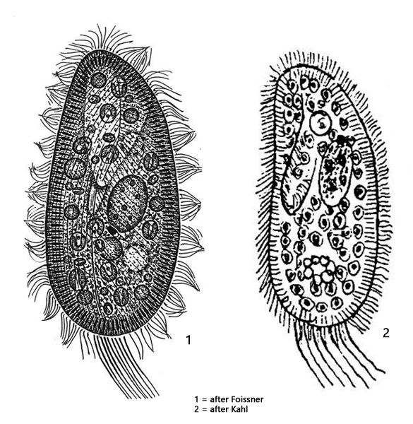
resting shape ellipsoidial to elongated ellipsoidal with a ventral depression
swimming shape cylindroidal, fast swimming
length 80–140 µm, width 37–61 µm
cytoplasm green due to symbiotic algae (about 500 cells)
macronucleus ellipsoidal, 20 x 14 µm
micronucleus compact, 2.4 x 5.7 µm with a hyaline cap
two contractile vacuoles with collecting vesicles
each contractile vacuole with one excretion porus
oral opening near mid-body with pre-oral depression
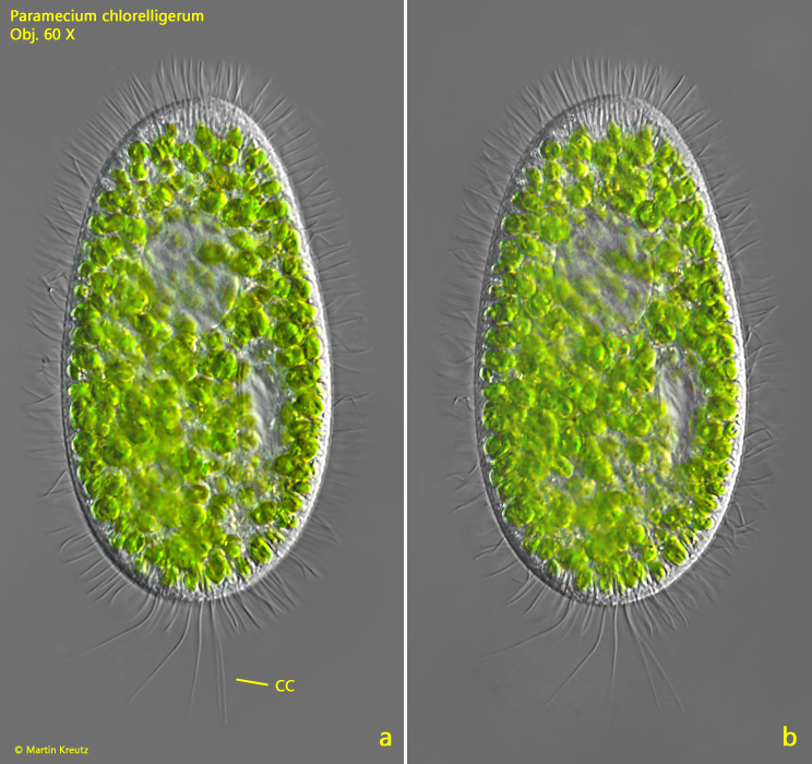
tuft of caudal cilia, 29 µm long on average
Paramecium chlorelligerum
In April 2010, I noticed a green Paramecium in samples from the Simmelried, which was very similar to Paramecium bursaria. However, there were a few peculiarities. The species swam extremely fast, taking on a cylindrical shape and had a tuft of unusually long caudal cilia. It turned out to be Paramecium chlorelligerum, which was first described by Kahl. He found Paramecium chlorelligerum at only one location, a small, clear bog pond with little detritus. He describes this species in just a few lines in the addendum to his monograph. Thereafter, no further finds or descriptions of this species are available. The population in the Simmelried now provided the opportunity for Paramecium clorelligerum to be described again in detail by Foissner, Stöck and myself (Kreutz et al., 2012, s. Literature).
I first found Paramecium clorelligerum in the mire outlet and later also in pond 2 of the Simmelried. In contrast to Paramecium bursaria, the specimens were mainly found in the uppermost mud layer, especially between aggregates of algae.
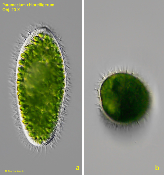
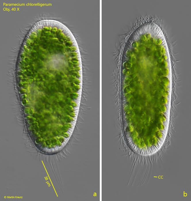
The most remarkable characteristic of Paramecium chlorelligerum is certainly its ability to take on a resting shape and a swimming shape. The resting shape is ellipsoidal with a ventral indentation in front of the central mouth opening (s. figs. 6 a-c and 7 a-f). However, the resting shape is not as pronouncedly slipper-like shaped as in Paramecium bursaria. This resting shape can very quickly change into a cylindroidal swimming shape, which is somewhat thickened in the apical third and circular in transverse view (s. figs. 4 a-b and 5 a-b). The swimming speed is very high at about 1 mm/sec. Paramecium chlorelligerum is the only ciliate I know that can actively take two forms.
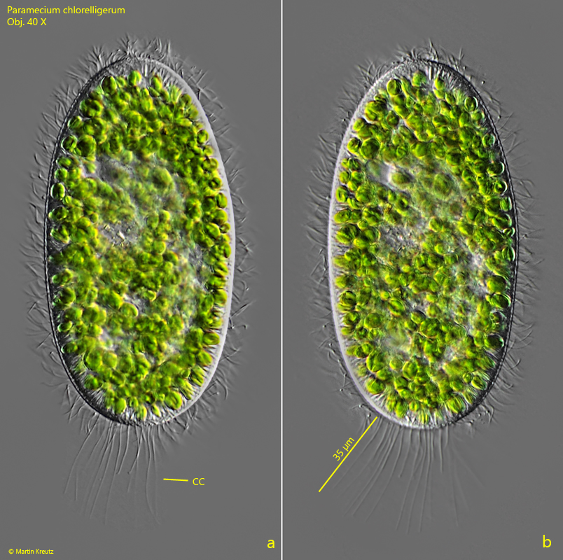
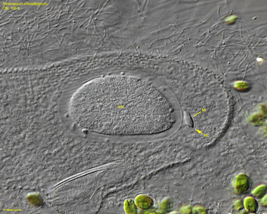
The conspicuous, long caudal cilia are on average 29 µm long (s. figs. 1 a-b, 2 a-b and 3 a-c). I have also observed specimens with caudal cilia 38 µm long (s. fig. 5 a), which corresponds to about one third of the body length. The two contractile vacuoles are surrounded by several collecting vesicles (s. fig. 11) and not by collecting ducts as in Paramecium bursaria. The excretory pores are located on the dorsal side. Each contractile vacuole has only one excretory pore (s. fig. 12). The macronucleus is oval with a small, highly refractive micronucleus, which has a hyaline cap (s. figs. 8 and 9). It sometimes appears wedge-shaped. The symbiotic algae, which fill the entire cell, are broad ellipsoidal (s. fig. 10). The cells are on average 7.7 X 5.5 µm in size and belong to the genus Meyerella. The closest related free-living species is Meyerella planktonica (99.1 % match of the 18S RNA gene sequence).
Fig. 1 a-b:Paramecium chlorelligerum. L = 123 µm. A freely swimming specimen from dorsal. Note the tuft of long caudal cilia (CC). Obj. 60 X.
Fig. 2 a-b:Paramecium chlorelligerum. L = 121 µm. A second, freely swimming specimen. The caudal cilia (CC) have a length of 35 µm. Obj. 60 X.
Fig. 3 a-c:Paramecium chlorelligerum. L = 113 µm. A third, freely swimming specimen from ventral. CC = tuft of caudal cilia. Obj. 60 X.
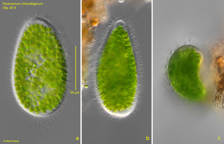
Fig. 4 a-b:Paramecium chlorelligerum. L = 135 µm. The cylindroidal swimming shape with a widened anterior third (a). In apical view (b) the swim-form is circular. Obj. 20 X.
Fig. 5 a-b:Paramecium chlorelligerum. L = 129 µm. Two different specimens that have take on the swimming shape. Obj. 20 X.
Fig. 6 a-c:Paramecium chlorelligerum. L = 113 µm. The broader, ellipsoidal resting shape from ventral (a), lateral (b) and apical (c). Note the ventral depression of the resting shape (c). Obj. 40 X.
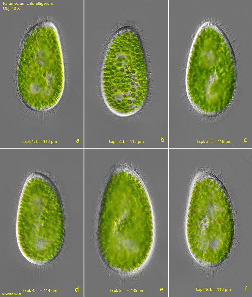
Fig. 7 a-f:Paramecium chlorelligerum. Six different specimens in the resting-form. Obj. 40 X.
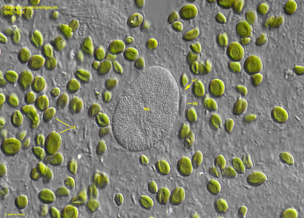
Fig. 8:Paramecium chlorelligerum. The macronucleus (Ma) and the adjacent micronucleus (Mi) in a strongly squashed specimen. Note the hyaline cap (HC) of the micronucleus. Obj. 100 X.
Fig. 9:Paramecium chlorelligerum. The macronucleus (Ma) and the micronucleus (Mi) in a second squashed specimen. HC = hyaline cap of the micronucleus, SA = symbiotic algae. Obj. 100 X.
Fig. 10:Paramecium chlorelligerum. The symbiotic algae are broad ellipsoidal with a length of 4.8–9.6 µm. They are members of the genus Meyerella. Obj. 100 X.
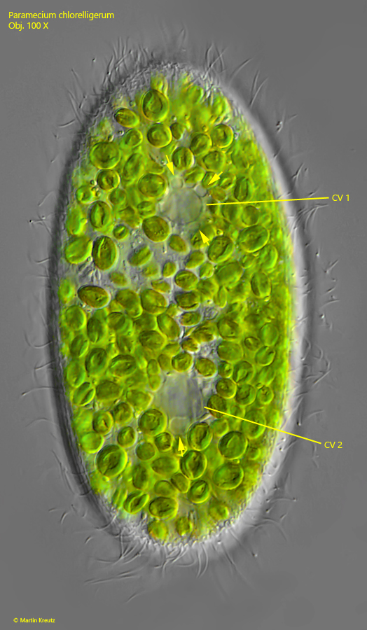
Fig. 11:Paramecium chlorelligerum. The two contractile vacuoles (CV 1, CV 2) are located dorsally. They are surrounded by several collecting vesicles (arrows). Obj. 100 X.
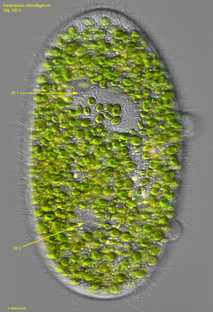
Fig. 12:Paramecium chlorelligerum. Each contractile vacuole has a single excretion pore (EP 1, EP 2). Obj. 100 X.
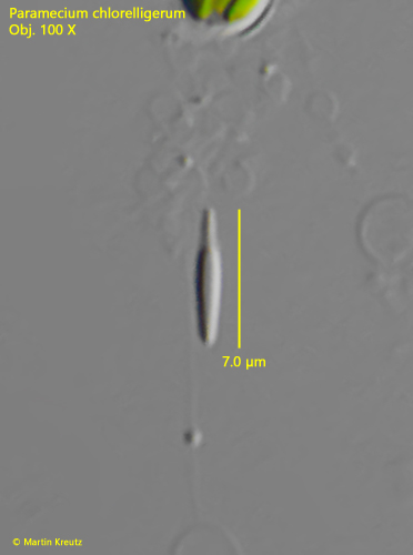
Fig. 13:Paramecium chlorelligerum. A resting extrusome with a length of 7 µm. Obj. 100 X.
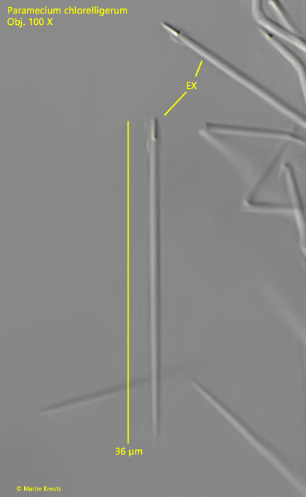
Fig. 14:Paramecium chlorelligerum. Exploded extrusomes (EX) with a length of 36 µm. Obj. 100 X.