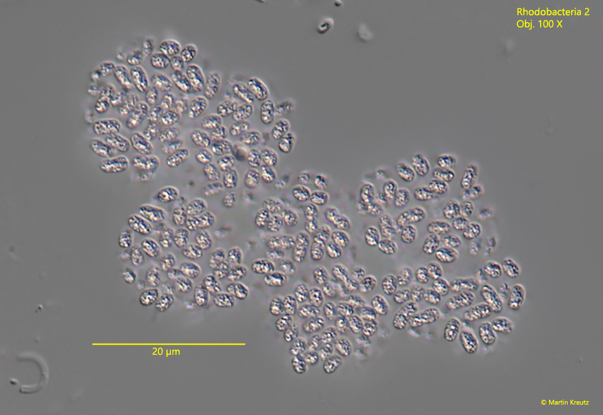
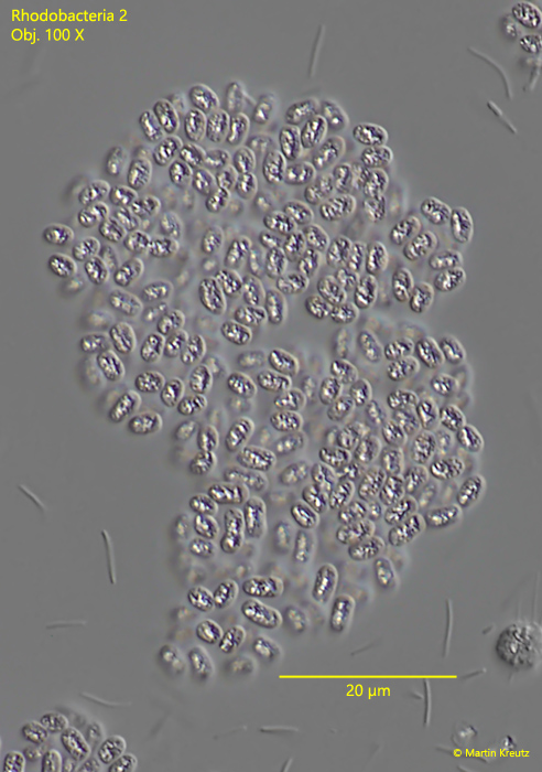
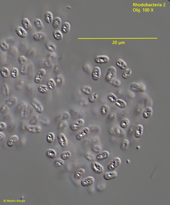
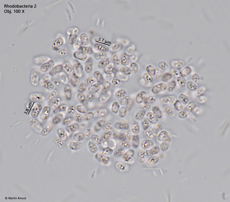
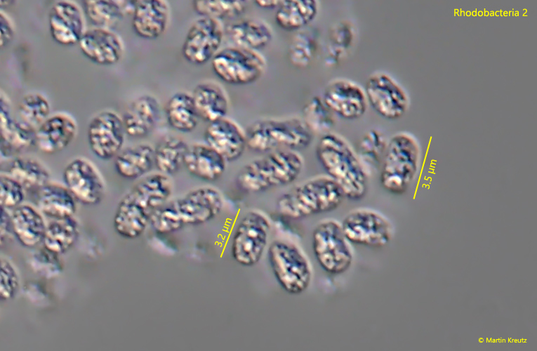
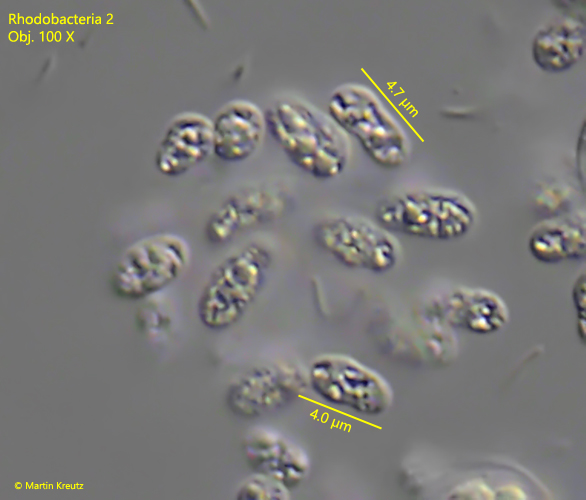
Rhodobacteria 2 Most likely ID: n.a. Synonym: n.a Sampling location: Simmelried Phylogenetic tree: n.a. Diagnosis: the cells are oval, sometimes irregularlylength 3 – 4.5 µmcells filled almost completely with irregularly shaped, highly refractive masscolorless or slightly pinkcolonies irregularly shaped without sharp outlineno visible gelatinuous sheatcolonies of about 30 – 70 µm in diameter No drawings from previous authors available. Fig. 1: Rhodobacteria 2. L = 3 – 4.5 µm. A slightly squashed colony. All cells are separated from each other. Obj. 100 X. Fig. 2: Rhodobacteria 2. L = 3 – 4.5 µm. A slightly squashed colony. Obj. 100 X. Fig. 3: Rhodobacteria 2. L = 3 – 4.5 µm. A third, slightly squashed colony. Obj. 100 X. Fig. 4: Rhodobacteria 2. L = 3 – 4.5 µm. A colony in brightfield. Obj. 100 X. Fig. 5: Rhodobacteria 2. L = 3 – 4.5 µm. The cells in detail. The cells are almost completely filled with a highly refractive mass. Obj. 100 X. Fig. 6: Rhodobacteria 2. L = 3 – 4.5 µm. The cells from a second colony in detail. Obj. 100 X. Download PDF