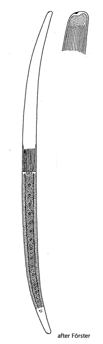
cell straight in middle part, ends attenuating and curved
length 400–770 µm width 25–35 µm
cell wall colorless or brownish
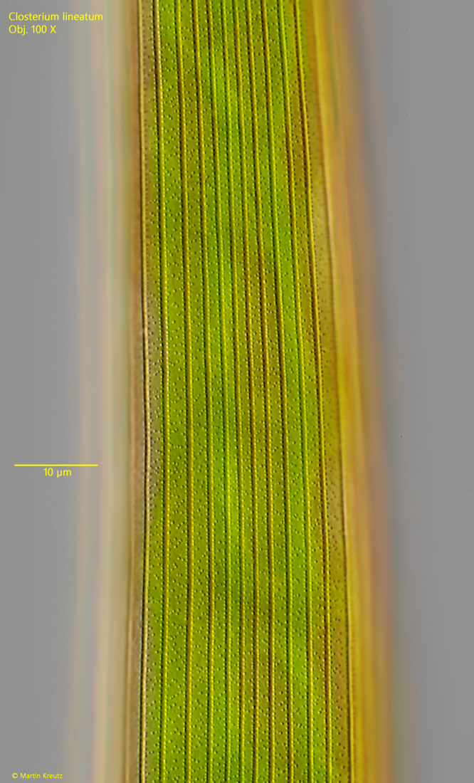
cell wall smooth or with distinct striae
puncta between striae
striae at the cell ends resolved into puncta
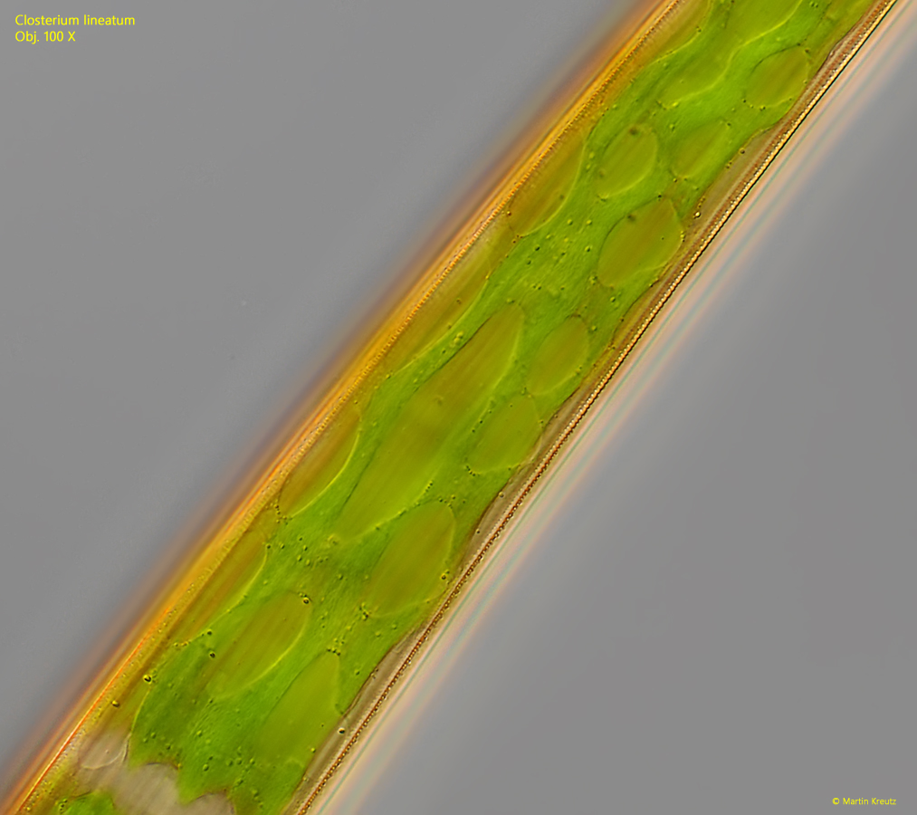
chloroplasts with 3–5 longitudinal ridges
9–22 pyrenoids in a row per semi-cell
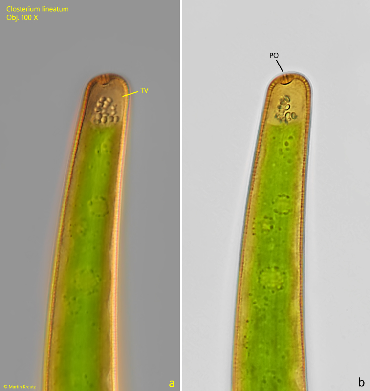
apices obliquely truncated with distinct apical pore
terminal vacuoles with each 4–10 crystals
girdle bands absent, sometimes pseudo girdle bands
Closterium lineatum
I found Closterium lineatum in the Simmelried until 1994, but not again after that.I found further specimens from the Ibmer Moor in Austria in 1995 and finally again from the Schwemm Moor (Austria) in 2025.
Closterium lineatum is one of the largest species in the genus Closterium.Most specimens are between 600 and 700 µm long.They are characterized by a straight middle section and then rapidly tapering and slightly curved ends.The apices are obliquely truncated and have a clearly visible, thickened pore (s. fig. 5 a-b).The striation of the cell wall was always very clear in my specimens (s. fig. 3).The cell wall is also clearly punctured between the striations.
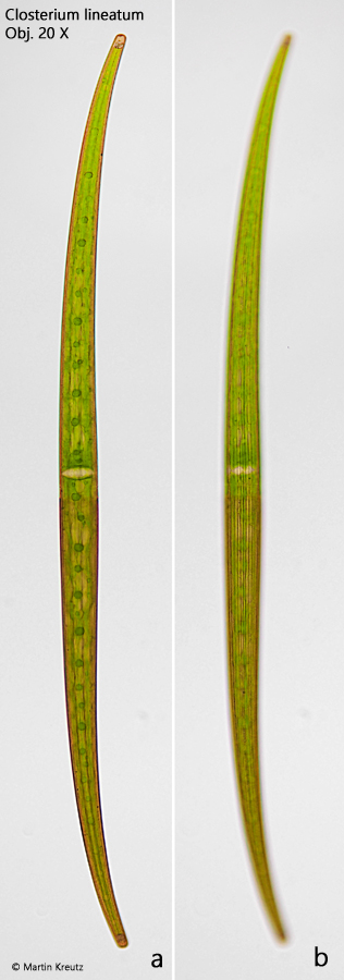
Fig. 1 a-b:Closterium lineatum. L = 765 µm. Two focal planes of a specimen in brightfield illumination. Obj. 100 X.
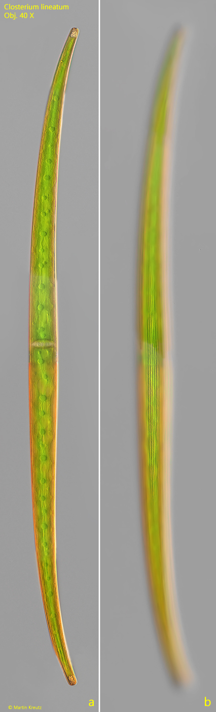
Fig. 2 a-b:Closterium lineatum. L = 765 µm. The same specimen as shown in fig. 1 a-b in DIC and at higher magnification. Obj. 100 X.
Fig. 3:Closterium lineatum. The striation of the cell wall in detail. Between the striae the cell wall is punctured. Obj. 100 X.
Fig. 4:Closterium lineatum. The chloroplast appears reticulate. Obj. 100 X.
Fig. 5 a-b:Closterium lineatum. The apex with a distinct, thickened pore (PO) in DIC (a) and in brightfield illumination (b). The terminal vacuole (TV) is filled with several gypsum crystals. Obj. 100 X.