So far, I have only found 3 specimens of Spathidiodes euglenivora. Two specimens came from the Purren Pond and one specimen from the Simmelried. I found all three specimens in the months of October and November.
Spathidiodes euglenivora was originally described by Kahl as Spathidiella euglenivora. Later, the species was transferred to the genus Spathidiodes by Foissner & Xu (2007). Foissner & Xu also mention that the species within the genus Spathidiodes (there are only 3) are all insufficiently studied, and it is questionable whether separation into a distinct genus is necessary.
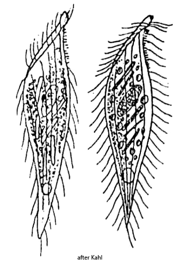
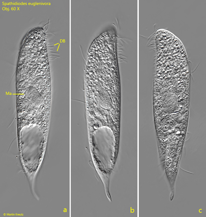
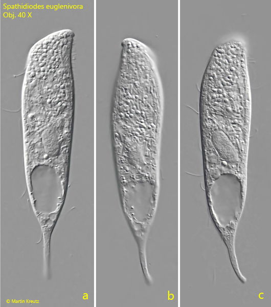
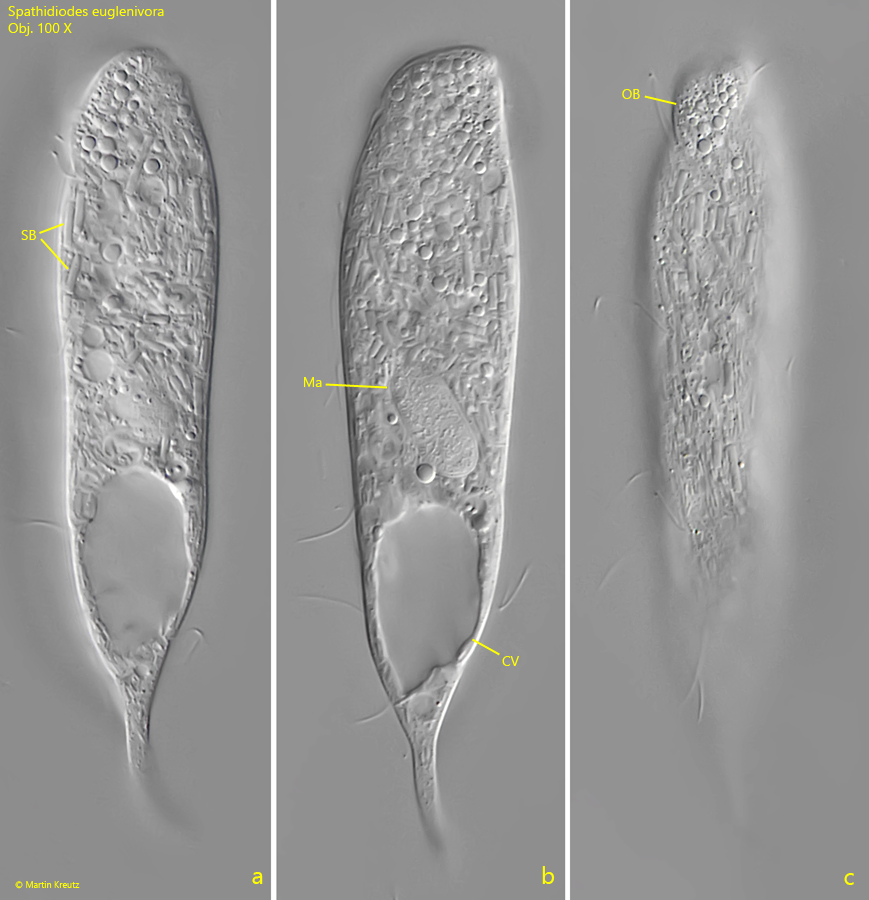
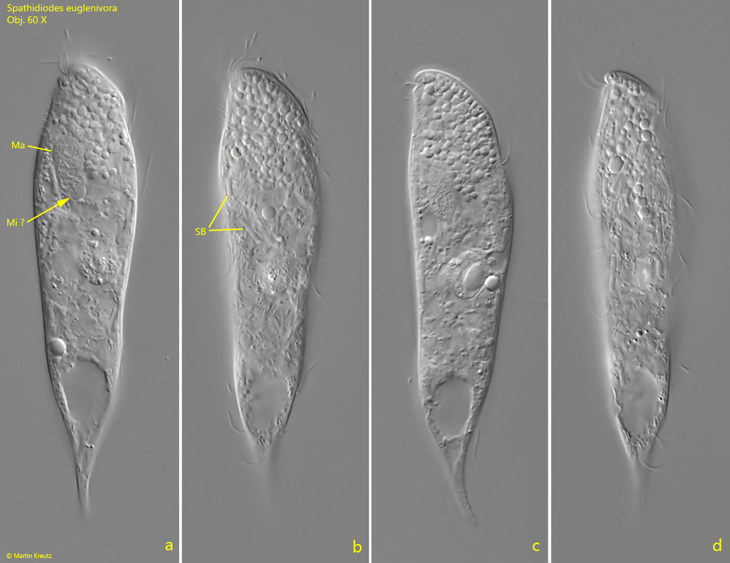
In fresh samples, the specimens of Spathidiodes euglenivora stand out due to their shape. The anterior two-thirds of the body are almost parallel-sided before the posterior third tapers into a tail-like end after the large, subterminal contractile vacuole. In my opinion, the drawings by Kahl (s. drawings above) do not represent this shape well. However, specimens of Spathidiodes euglenivora with the same shape as those in my population were also found by Silverman (2026) in the USA (s. link below).
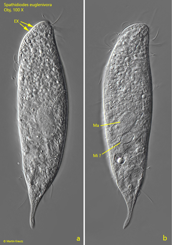
The oral bulge slopes down at an approximately 45° angle towards the ventral side. On the dorsal side, the oral lip is said to have a beak-like extension, which I could never see as clearly as Kahl depicted it (s. drawings above). Kahl mentions that no extrusomes are visible in the oral bulge. However, in at least one specimen from my population, I was able to observe rod-shaped extrusomes with a length of 2.5–3 µm (s. fig. 2 a). The dorsal brush was described by Kahl as short. In my specimens, however, the bristles were strikingly long (s. fig. 1 a). In all specimens, I was able to recognize the ellipsoidal macronucleus, but in no case could I clearly identify the micronucleus.
Kahl described Spathidiodes euglenivora as a food specialist that feeds exclusively on one species of a colorless euglenid. He was able to detect numerous paramylon grains of these euglenids in the cytoplasm. However, in my specimens, I could not detect paramylon grains in any case. Instead, the cytoplasm was filled with lipid droplets and numerous symbiotic bacteria (s. fig. 4 a-c). These symbiotic bacteria are not mentioned by either Kahl or Foissner & XU. This may be because they can only be seen at high magnification between the lipid droplets. As far as I could recognize it, at least two types of symbiotic bacteria are present, which bear a strong resemblance to those in Discomorphella pectinata.
More images and information on Spathidiodes euglenivora: Jeffrey Silverman-iNaturalist-Spathidiodes euglenivora