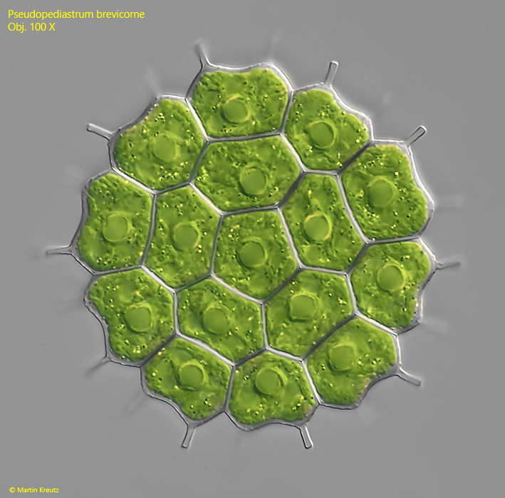
So far, I have found Pseudopediastrum brevicorne exclusively in the plankton of Lake Constance. However, the species occurs there only very rarely. The photos shown below are from a specimen I found in September 2022.
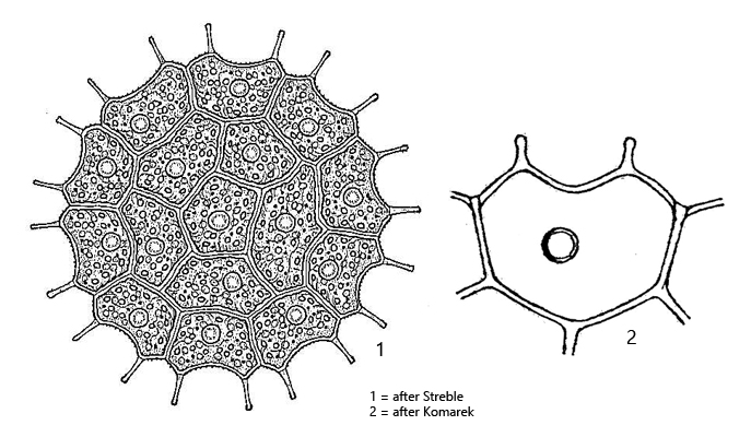
Pseudopediastrum brevicorne can be recognized by the short, hyaline lobes, which are sometimes slightly knob-shaped at the distal end, and by the shallow indentation between the lobes. In addition, there are no intercellular spaces between the cells.
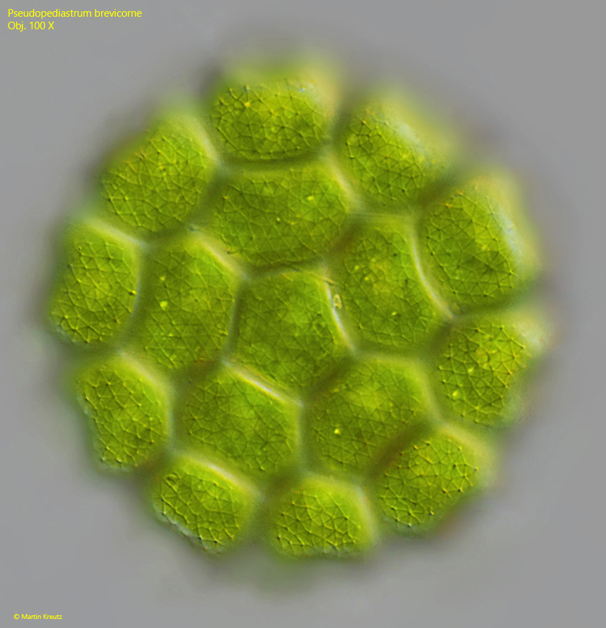
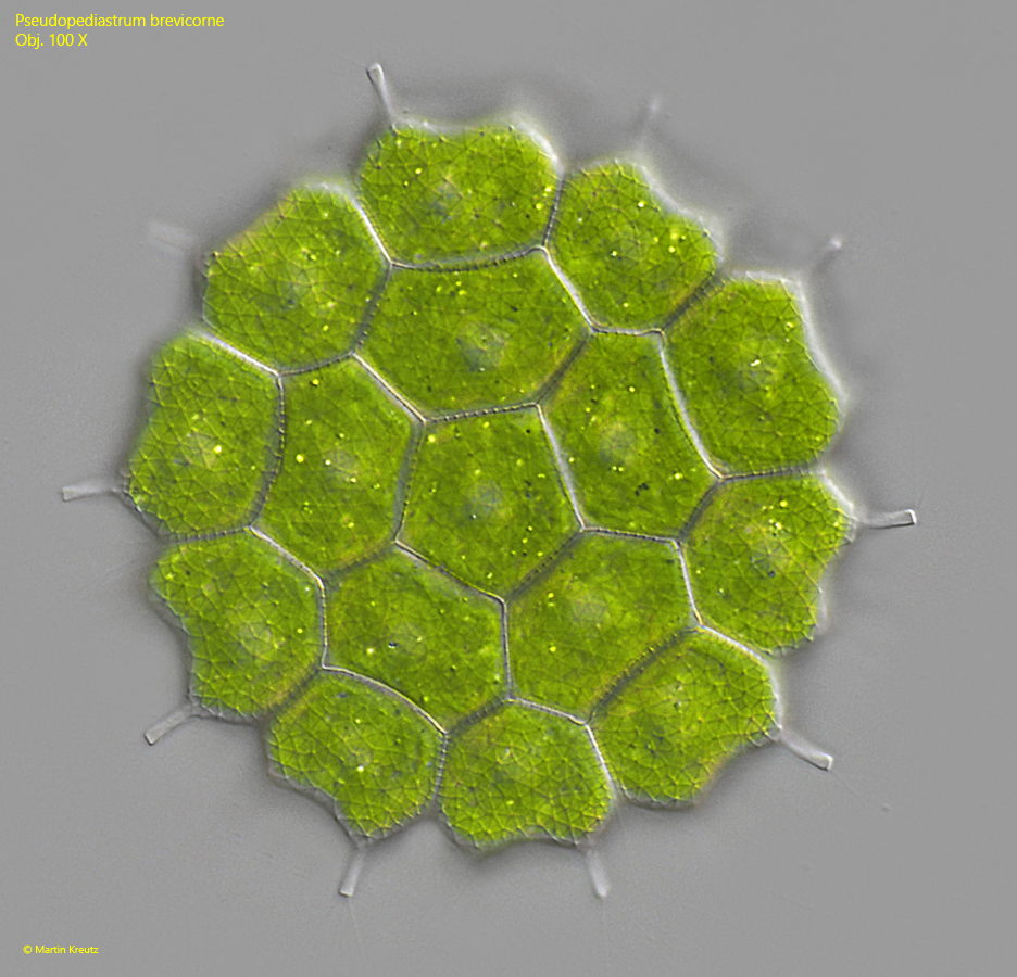
My specimen had a diameter of 76 µm and 16 cells (s. fig. 1). This is significantly larger than described by Komárek & Jankovská (2001), who reported a maximum of 40 µm. The same authors described the cell wall as slightly punctate. However, the specimen I found had a delicate but clearly visible reticulate structure (s. figs. 2 and 3). So far, I have not found a second specimen to verify the consistency of this characteristic.
Fig. 1:Pseudopediastrum brevicorne. D = 76 µm. A coenobium with 16 cells. Obj. 100 X.
Fig. 2:Pseudopediastrum brevicorne. D = 76 µm. The cell wall of the specimen as shown in fig. 1 has a delicate reticulate pattern. Obj. 100 X.
Fig. 3:Pseudopediastrum brevicorne. D = 76 µm. The slightly squashes specimen as shown in fig. 1 with focal plane on the reticulate pattern of the cell wall. Obj. 100 X.