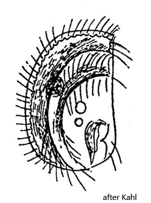
right side flat with 3 rows of cilia interrupted in middle
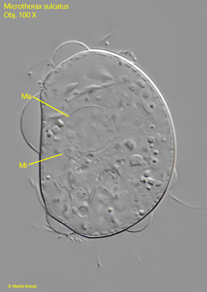
macronucleus spherical in mid-body
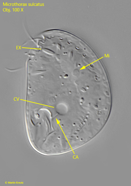
contractile vacuole near mouth opening
extrusomes spindle shaped
oral apparatus at posterior end of cell on right side
Microthorax sulcatus
In August 2026, I found a population of Microthorax sulcatus in samples from the pond of the waste disposal company Constance. These were not plankton samples, but rather some stems and leaves of yellow water lily (Nuphar lutea), which grows in large numbers in this pond. After a few days, the plant parts began to rot, and a strong mass development of Paramecium caudatum and Paramecium aurelia occurred. Many specimens of Microthorax sulcatus were found among these Paramecia. However, the population of Microthorax sulcatus collapsed again after only a few days.
Microthorax sulcatus can be recognized by its distinctly semi-circular body. The ventral side is almost straight or only slightly concave, while the dorsal body margin is semi-circular, sometimes somewhat asymmetrically shifted dorsally. On the right side, the mouth opening is located at the posterior end, and three rows of cilia run along this side, following the shape of the dorsal margin. In the middle of the body, all of these ciliary rows are interrupted (s. fig. 2 b).
The left side of the body was described by neither Engelmann, Penard, nor Kahl. I too was unable to examine it in greater detail, since most specimens were oriented under the coverslip with their left side facing the slide. The other characteristics are typical of the genus Microthorax. The round macronucleus lies approximately in the middle of the body together with a micronucleus (s. fig. 4). The contractile vacuole is located at the level of the mouth opening and is connected to it by a short canal (s. fig. 3). The extrusomes are spindle-shaped. In Microthorax sulcatus, according to my measurements, they are 4.5–4.8 µm long (s. fig. 5).
Engelmann (1862) also described the similar species Microthorax pusillus, which likewise has a semicircular body. However, this species is smaller, measuring 20–30 µm, which is why I believe that Microthorax sulcatus is present in my population. All specimens I examined were longer than 30 µm.
Fig. 1 a-f:Microthorax sulcatus. L = 38 µm. Different focal planes of a slightly squashed specimen from right. Obj. 100 X.
Fig. 2 a-c:Microthorax sulcatus. L = 35 µm. A second specimen from right. Note the rows of cilia (CR1–CR3) interrupted in mid-body. Ma = macronucleus, MO = mouth opening. Obj. 100 X.
Fig. 3:Microthorax sulcatus. L = 34 µm. A slightly squashed specimen from left. Note the short canal (CA) between the contractile vacuole (CV) and the mouth opening. EX = extrusomes, Mi = micronucleus. Obj. 100 X.
Fig. 4:Microthorax sulcatus. L = 38 µm. The spherical macronucleus (Ma) with the adjacent micronucleus (Mi) in a squashed specimen. Obj. 100 X.
Fig. 5:Microthorax sulcatus. The extrusomes (EX) are spindle-shaped with a length of 4.5–4.8 µm. Ma = macronucleus. Obj. 100 X.