Sonderiella 2
Most likely ID: Sonderiella nov. spec.
Synonym: n.a.
Sampling location: Simmelried, Purren pond
Phylogenetic tree: Sonderiella
Diagnosis:
- body broad ellipsoid, laterally flattened
- left side flat, right side slightly domed
- three longitudinal ribs on left and right side
- length 19–23 µm
- mouth opening ventral near anterior end
- lower lip sickle-shaped protrudes ventral side
- upper lip U-shaped
- ventral field of cilia, parallel rows
- ventral two elongated cilia at posterior third
- contractile vacuole ventral, below mouth opening
- pellicle covered with layer of symbiotic bacteria
- macronucleus ellipsoid, near anterior end
- posterior an irregular mass of symbiotic bacteria
No drawings from previous autors available.
In August and October 2006 I found the first specimens of a ciliate in the Simmelried, which I consider, based on its characteristics, to belong to the genus Sonderiella. In October 2017 I found more specimens in the Simmelried and finally in February 2002 also some specimens in the Purren pond. All specimens were found in a layer of decomposed leaves.
The genus Sonderiella, established by Kahl in 1928, is distinguished by a special structure of the oral apparatus. It consists of a sickle-shaped lower lip that rises clearly above the flat ventral side. Opposite this is an upper, U-shaped lip that is significantly smaller. Between the lips a depression leads to the left into the pharynx. Only a single species, Sonderiella scandens, was described by Kahl. So far there has been only one redescription of Sonderiella scandens by Jeffrey Silverman from July 2025 (s. Sonderiella scandens, iNaturalist).
In addition to the adherent symbiotic bacteria on the pellicle, Sonderiella 2 also has an irregularly shaped conglomerate of densely packed symbiotic bacteria in the cytoplasm, which fills almost the entire posterior third of the body (s. figs. 1 d and 2 c). The function of these symbiotic bacteria can only be speculated upon. The macronucleus of Sonderiella 2 has low contrast and is located near the anterior end (s. fig. 1 b). As far as I was able to observe, there is probably only a single micronucleus, which is attached to the macronucleus (s. fig. 1 b). The contractile vacuole lies ventrally beneath the oral aperture (s. fig. 1 d). The oral aperture consists of a rigid lower lip and a movable upper lip (s. figs 2 b and 3 a). The lower lip rises above the flat ventral side. This type of oral apparatus probably serves to scrape bacteria from the substrate.
According to my research, no ciliate with such a combination of characteristics has been described either within the genus Sonderiella or in any other genus, so it is probably a hitherto undescribed species, Sonderiella nov. spec.
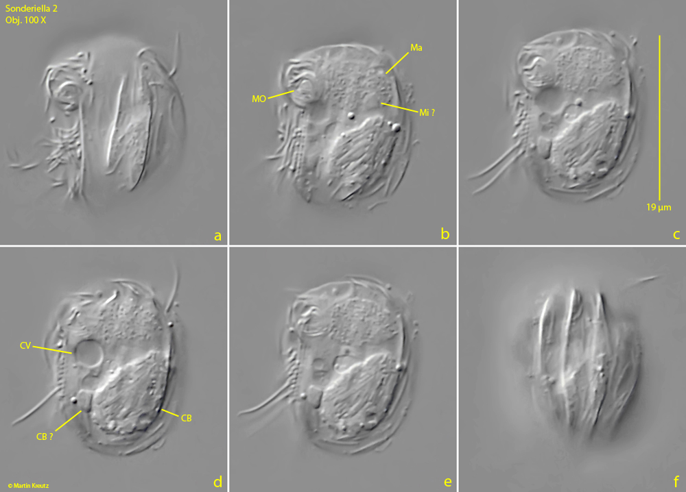
Fig. 1 a-f: Sonderiella 2. L = 19 µm. Different focal planes of a specimen found in February 2002 in the Purren pond from the left side (a) to the right side (f). On both sides of the body each 3 longitudinal ribs are visible. The posterior third of the body is filled with an irregular shaped conglomerate of densely packed symbiotic bacteria (CB, CB ?). Near the anterior end the macronucleu (Ma) is located. CV = contractile vacuole, Mi ? = probably the micronucleus, MO = mouth opening. Obj. 100 X.
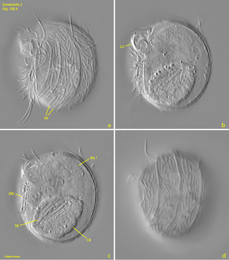
Fig. 2 a-d: Sonderiella 2. Different focal planes of a slightly squashed specimen from the left side (a) to the right side (d). The pellicle is completely covered with rod-shaped, symbiotic bacteria (SB). The lower lip (LLI) of the oral apparatus protrudes the ventral side. Below the mouth opening the ventral side is covered with a parallel rows of cilia (VRC). CB = conglomerate of symbiotic bacteria, Ma = probably the macronucleus. Obj. 100 X.

Fig. 3 a-d: Sonderiella 2. L = 23 µm. A freely swimming specimen found in October 2017 in the Simmelried from left (a, c), ventral (b) and apical (d). The lower lip (LLI) and the upper lip (ULI) of the oral apparatus are visible. VFC = ventral field of cilia. Obj. 100 X.