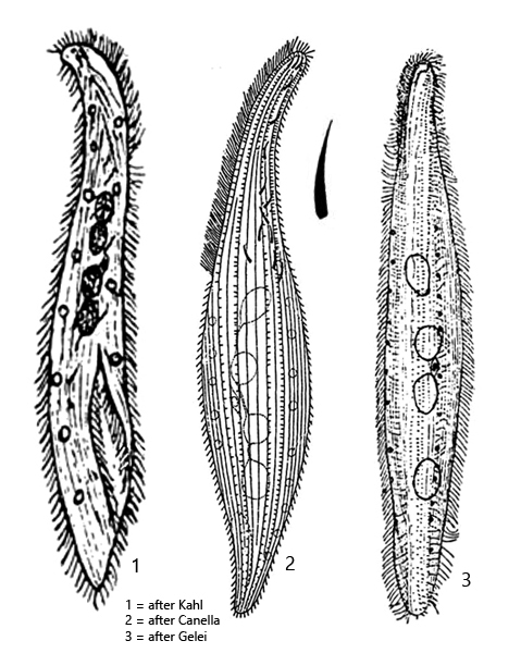
body slender laceolate, slightly sigmoid, laterally flattened
length 200–360 µm, width 50–60 µm
mouth slit 1/4–1/3 of body length (visible when feeding)
right side with about 45 rows of cilia
left side with about 5 rows of short bristles
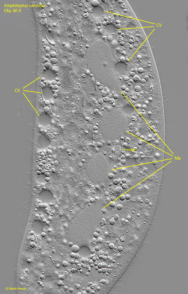
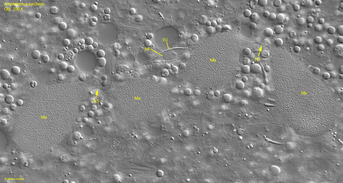
moniliform macronucleus consists of 4 nodules
in middle of nodules a spherical micronucleus in funiculus
one (sometimes two) rows of several contractile vacuoles on ventral and dorsal margin
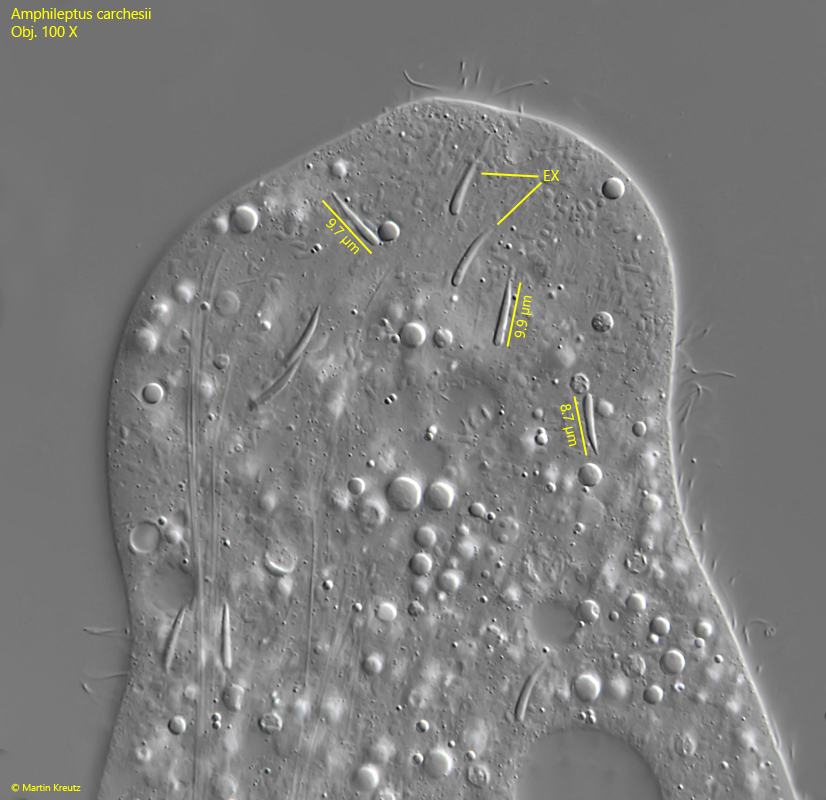
extrusomes tusk-shaped, about 11 µm long
extrusomes attached to anterior end of oral slit
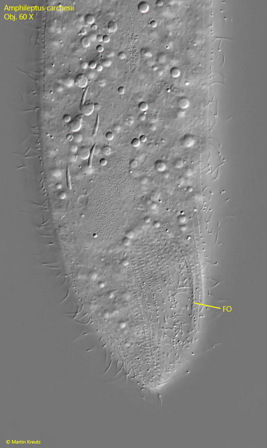
posterior on right side a distinct groove (called fossa)
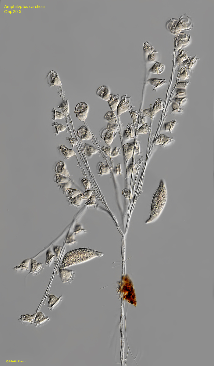
parasitizes colonies of peritrich ciliates (e.g. Carchesium)
attached to the host colonies via a thread
Amphileptus carchesii
So far, I have only found Amphileptus carchesii once on a colony of the peritrich ciliate Carchesium polypinum. Amphileptus carchesii parasitizes colonies of Carchesium polypinum and other peritrich ciliates (e.g., Vorticella sp.). In this case, the infested colony was on a submerged slide on which growth had developed within 9 days.
There are still very few records of this rare species. As far as I know, detailed descriptions are only available from Schneider (1988) and Foissner et al. (1992). The specimens are almost never found free-swimming, but mostly on colonies of the peritrich ciliate Carchesium polypinum. On these colonies, Amphileptus carchesii lives and feeds on the zooids. Each specimen of Amphileptus carchesii ingests about 40 zooids per day (Foissner et al., 1992). In doing so, Amphileptus carchesii always attacks the oral apparatus of the zooids with its extrusomes. The zooids, which are then paralyzed, are swallowed whole. Well-nourished specimens eventually form cysts, which can be found attached to the stalks of the colony.
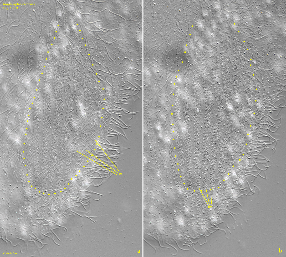
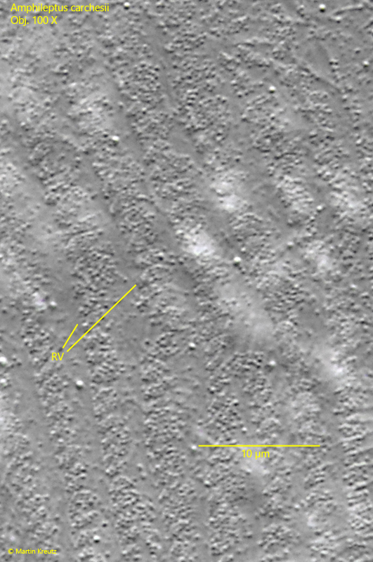
Amphileptus carchesii is extremely well adapted to a parasitic life on colony-forming, peritrichous ciliates. In order to never lose contact with the host colony, Amphileptus carchesii forms a thread at its posterior end, with which the individuals attach themselves to the stalks of the colony (s. fig. 3). The thread is lengthened with every movement, so that the stalks of infested colonies are often wrapped in many threads. The thread is formed in a groove at the posterior end on the right side. The groove is called the fossa. Exactly how the thread is formed is not clear. Under light microscopy, it can be seen that the fossa is lined with parallel rows of bristle-like short cilia. Between these rows of cilia, just beneath the pellicle, there are rows of densely packed vesicles (s. figs. 7 a-b and 8). Possibly, the contents of these vesicles provide the components to form the thread.
Amphileptus carchesii can easily be confused with the similar species Amphileptus claparedii, which also parasitizes colonies of peritrichous ciliates, especially Carchesium polypinum. However, Amphileptus claparedii does not possess a fossa and only has two macronuclear nodules. Furthermore Amphileptus claparadii lacks extrusomes.
Fig. 1:Amphileptus carchesii. L = 242 µm + 220 µm. Two specimen parasitize a colony of Carchesium polypinum. One specimen feeds on the zooids, while the other is freely swimming around the colony. Obj. 20 X.
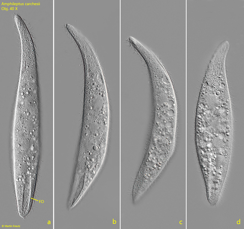
Fig. 2 a-d:Amphileptus carchesii. L = 304 µm. A freely swimming specimen from right. Note the groove at the posterior end called fossa (FO). Obj. 40 X.
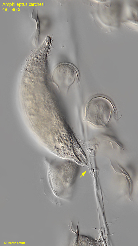
Fig. 3:Amphileptus carchesii. L = 226 µm. A specimen that eats the zooids of Carchesium polypinum is connected to the colony by a thread (arrow) to prevent detachment and drifting away. The thread is formed by the fossa. Obj. 40 X.
Fig. 4:Amphileptus carchesii. The nuclear apparatus in a squashed specimen. It consists of 4 macronuclear nodules (Ma) and a round micronucleus in the center (Mi). On the ventral side as well as on the dorsal side rows of contractile vacuoles (CV) are visible. Obj. 60 X.
Fig. 5:Amphileptus carchesii. The nuclear apparatus in detail. The macronuclear nodules (Ma) are connected via thin strands (NS). The spherical micronucleus (Mi) is enclosed in a case called funiculus (FU). Obj. 100 X.
Fig. 6:Amphileptus carchesii. The fossa (FO) at the posterior end on the right side in a slightly squashed specimen. Obj. 60 X.
Fig. 7 a-b:Amphileptus carchesii. Two focal planes of the fossa in a squashed specimen. The fossa (marked by a dotted line) is lined with parallel rows of short, bristle-like cilia (RC, a). Directly beneath the pellicle, rows of accumulations of small vesicles are visible (RV, b), which may possibly contain the components for the construction of the thread. Obj. 100 X.
Fig. 8:Amphileptus carchesii. A crop from fig. 7 b to show the rows of vesicles (RV) under the pellicle of the fossa in detail. Obj. 100 X.
Fig. 9:Amphileptus carchesii. The tusk-shaped extrusomes (EX) at the anterior end in a strongly squashed specimen. The extrusomes have a length of 6.6–9.7 µm. Obj. 100 X.