stomach yellow or yellow greenish due to ingested algae
toes about 35 µm long, slightly dorsally curved
No drawings from previous autors available.
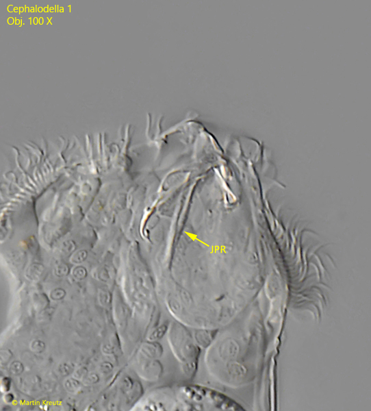
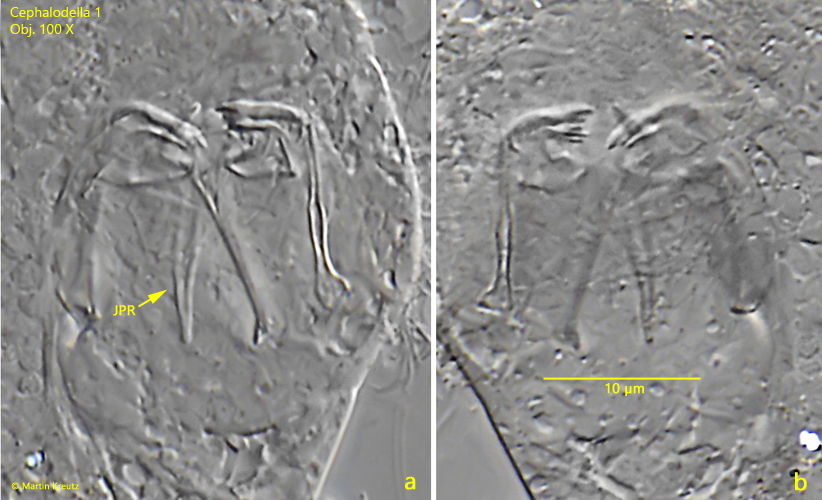
In July 2024 I found a few specimens of Cephalodella 1 in the pond of the convent Hegne. At first I thought this species was Cephalodella gibboides (Wulfert, 1950, s. fig. 6), because in the trophi of Cephalodella 1 I could observe Y-shaped connected pleural rods (s. figs. 4 and 5 a-b). This feature is possessed only by Cephalodella wrighti (Wulfert, 1960) and Cephalodella gibboides. Since Cephalodella wrighti has only very short toes, my assumption was that my specimens are Cephalodella gibboides. However, Cephalodella 1 shows some essential differences from Cephalodella gibboides.
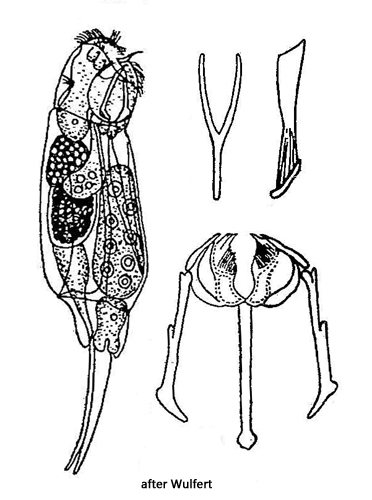
The specimens of Cephalodella 1 were between 120–140 µm long and thus about 30% smaller than Cephalodella gibboides (184–206 µm). In addition, my specimens lacked the double frontal eye. The gastric glands of Cephalodella gibboides are filled with fat globules, which was also not the case in my specimens.
Since there is therefore no alternative among the previously known species with Y-shaped pleural rods, Cephalodella 1 could be a species not yet described.
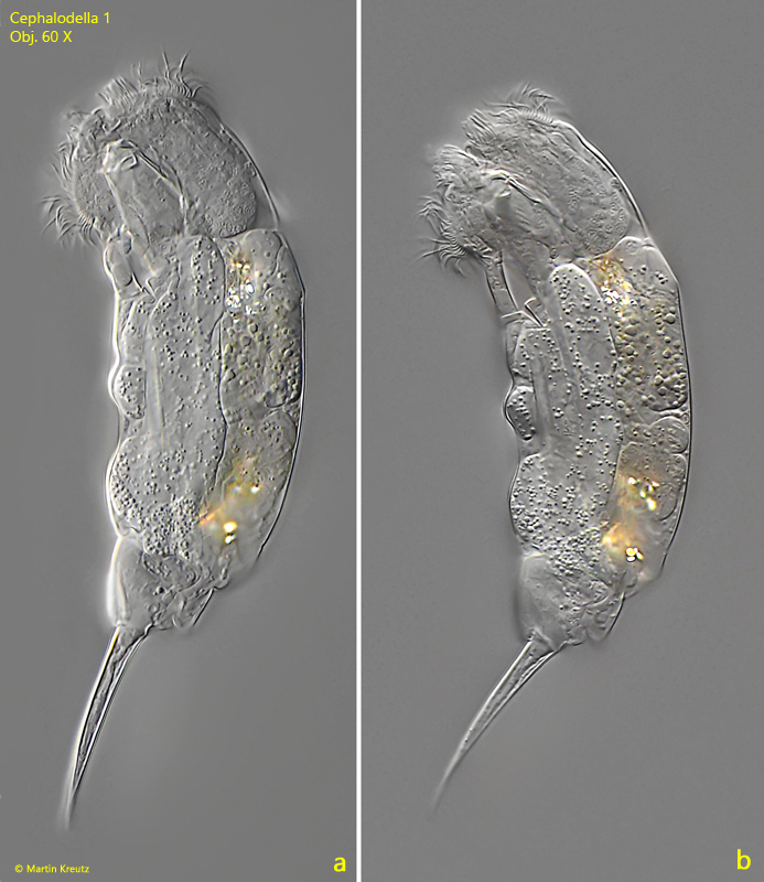
Fig. 1 a-b: Cephalodella 1. L = 128 µm. A freely swimming specimen from left. Obj. 60 X.
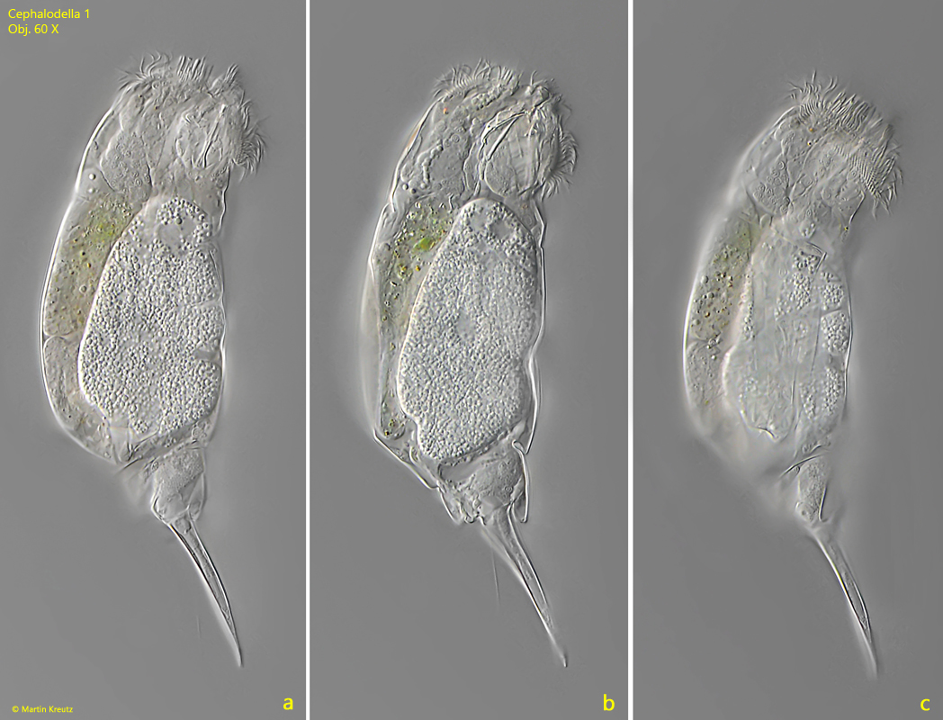
Fig. 2 a-c:Cephalodella 1. L = 125 µm. A second freely swimming specimen from right. Obj. 60 X.
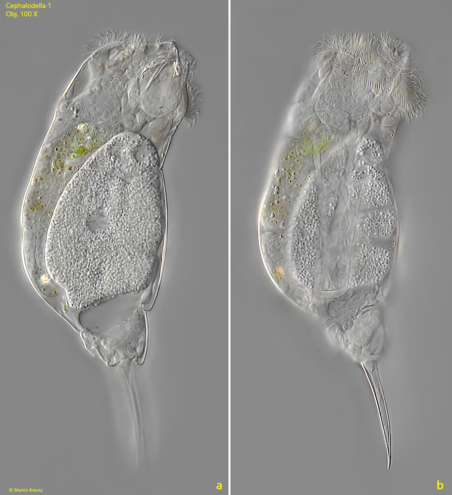
Fig. 3 a-b:Cephalodella 1. L = 125 µm. The same specimen as shown in fig. 2 a-c slightly squashed. Obj. 100 X.
Fig. 4:Cephalodella 1. The head of the specimen as shown in fig. 3 a-b with focal plane on the trophi. Note the Y-shaped, joined pleural rods. Obj. 100 X.
Fig. 5 a-b:Cephalodella 1. Two focal planes of the trophi in a squashed specimen. JPR = joined pleural rods. Obj. 100 X.
Fig. 6: Drawing of the similar species Cephalodella gibboides after Wulfert.