terminal vacuoles with single or few small crystals
nucleus central
Closterium incurvum
I find Closterium incurvum quite frequently in the Simmelried, but always only sporadically. I have not yet been able to find this species in my other sampling sites. The most important distinguishing feature is the small size of less than 80 µm and the strongly curved shape, which can almost form a semi-circle.
In my population there was always only 1 pyrenoid per half cell. I have never found cells with more pyrenoids, although according to Förster (1982) there can be up to 3 pyrenoids per half cell.
The differentiation from the very similar species Closterium venus is very difficult. The cells of this species are larger (up to 100 µm) and less curved. The semi-cells of Closterium venus usually contain more than one pyrenoid, but not always. Otherwise the characteristics are identical. Since the cells of my population are strongly curved and have a length of about 55–70 µm, they can be assigned to Closterium incurvum.
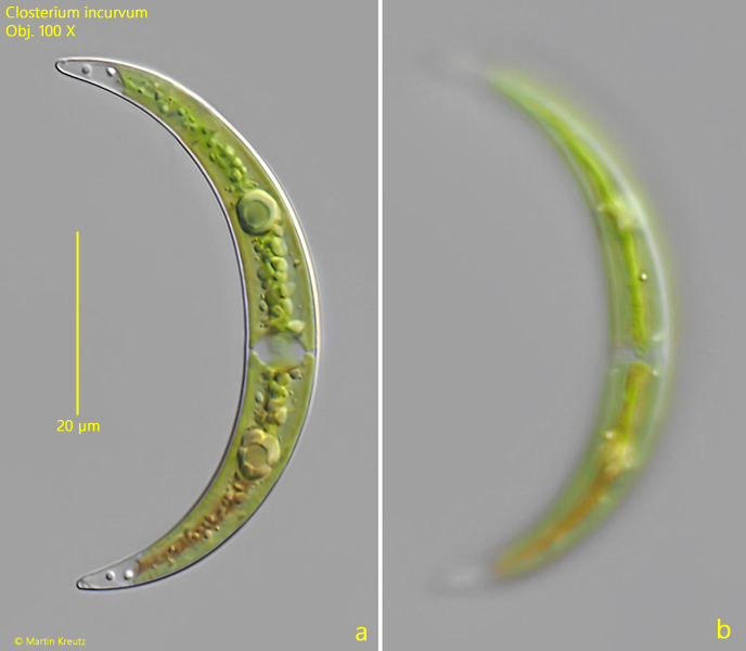
Fig. 1 a-b:Closterium incurvum. L = 64 µm. Two focal planes of a specimen in DIC. Note the small pores (PO) in the apices and the mooth cell wall without striation (b). Obj. 100 X.
Fig. 2 a-b:Closterium incurvum. L = 64 µm. The same specimen as shown in fig. 1 a-b in brightfield illumination. Obj. 100 X.
Fig. 3 a-b:Closterium incurvum. L = 58 µm. A second specimen found in the Schwemm Moor in July 2025. Obj. 100 X.