I find Colpoda maupasi exclusively in moss samples that I pour rainwater over in Petri dishes. After a few days, populations of varying sizes form.
Colpoda maupasi can be recognized mainly by the fact that the mouth opening is located at the border of the apical third of the body and is very small. In addition, Colpoda maupasi lacks a “beard” caused by elongated cilia of the mouth cilia, as is the case with Colpoda steinii, for example.
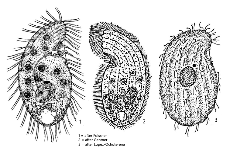
The specimens in my population were 38–60 µm long. The size can vary greatly depending on the food supply. Well-fed specimens are often pear-shaped. The keel above the mouth opening always had 5 ribs in my specimens (s. fig. 4), as described by Foissner (1993). The nucleolus in my specimens had the shape of divided clumps distributed throughout the macronucleus (s. fig. 4). The micronucleus was lenticular and located close to the macronucleus (s. fig. 4). The contractile vacuole is terminal.
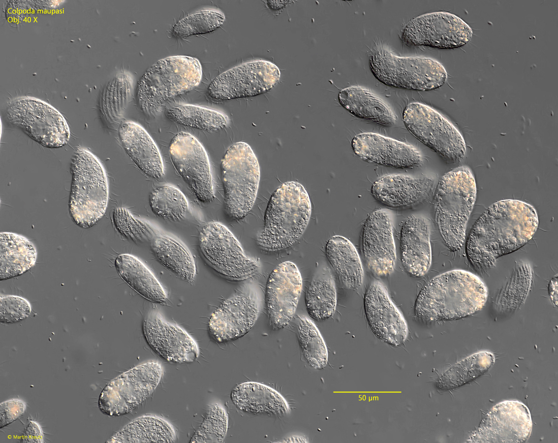
Fig. 1:Colpoda maupasi. L = 38–58 µm. An agglomeration of several specimens in a sample from wettened moss. Obj. 40 X.
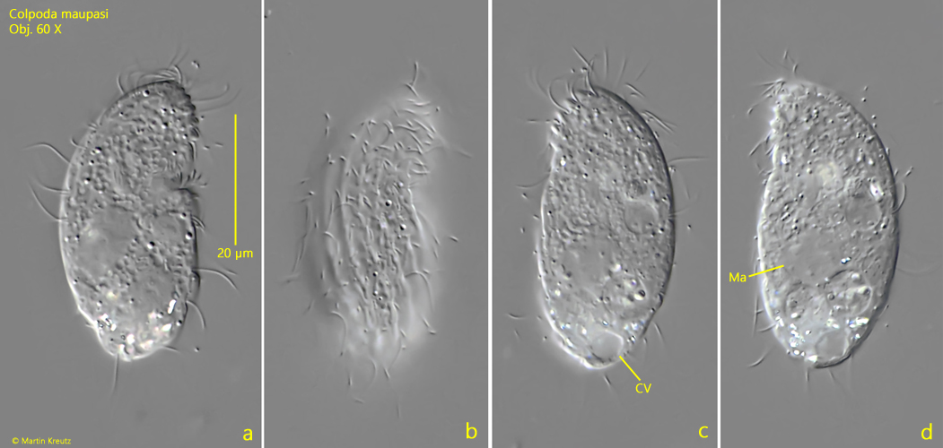
Fig. 2 a-d:Colpoda maupasi. L = 45 µm. A freely specimen from right (a, b) and from left (c, d). Obj. 60 X.
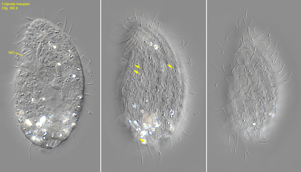
Fig. 3 a-c:Colpoda maupasi. L = 49 µm. A slightly squashed specimen from left. Note the mouth opening in the anterior third and the paired cilia of the somatic ciliature (arrows). Obj. 100 X.
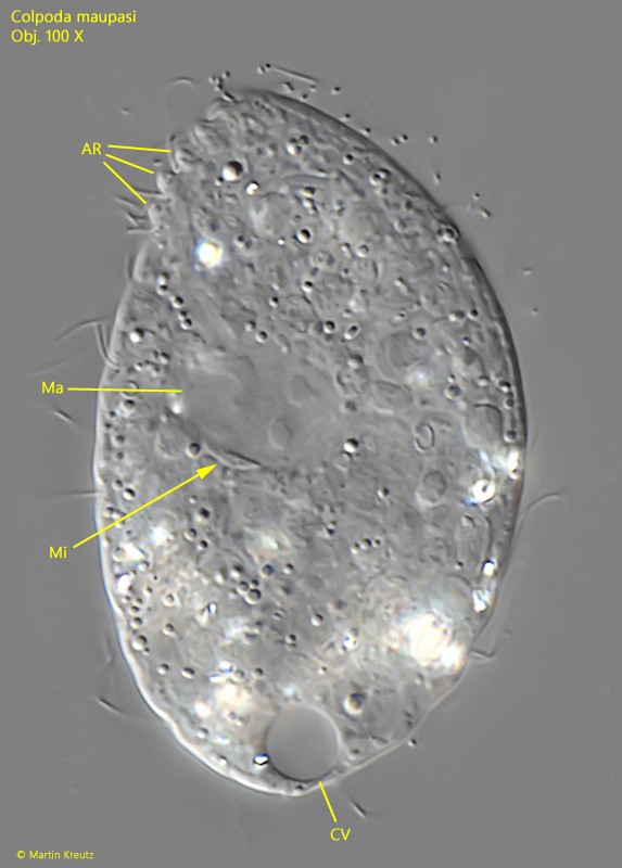
Fig. 4:Colpoda maupasi. A squashed specimen from left. In the anterior third 5 apical ribs (AR) are visible. CV = contractile vacuole, Ma = macronnucleus, Mi = micronucleus. Obj. 100 X.