pharynx with ejectisomes reaches mid-body or posterior third
two Maupas’ bodies present
pyrenoids absent
two chromatophores, brownish-greenish or olive
two flagella
contractile vacuole apical, near pharynx
Cryptomonas obovata
So far, I have only found Cryptomonas obovata in the Simmelried. The specimens are mainly found in floating or decomposing plant matter.
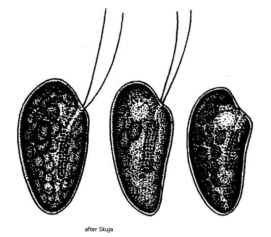
Cryptomonas obovata has only been described in detail by Skuja, who frequently found the species in Latvia and in the lakes of Uppland. A characteristic feature of this species is its approximately obovate shape with an oblique apical end. The apical end has no indentation, as is the case with Cryptomonas erosa. The dorsal side is always distinctly convex, while the ventral side is less convex or may even be concave (s. drawings above). There are always two Maupas’ bodies, which glow brightly in the DIC.
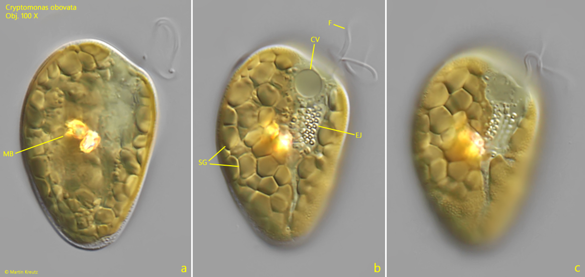
Skuja explicitly mentions the numerous starch grains, which are densely packed directly below the two chloroplasts. I can confirm this observation (s. fig. 2 b). The specimens in my population were between 45–55 µm long and thus slightly larger than those reported by Skuja. The chloroplasts in my specimens were brownish-yellow in color. A greenish tinge was only faintly perceptible.
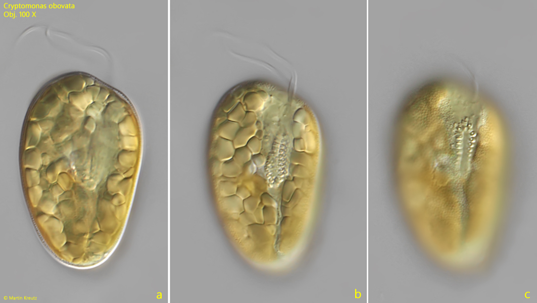
Fig. 1 a-c:Cryptomonas obovata. L = 50 µm. Three focal planes of a freely swimming specimen from right. Obj. 100 X.
Fig. 2 a-c:Cryptomonas obovata. L = 45 µm. Three focal planes of a second specimen from right. Note the densely packed starch grains (SG) located beneath the chloroplasts. CV = contractile vacuole, EJ = ejectisomes, F = flagellum, MB = Maupas’ bodies. Obj. 100 X.
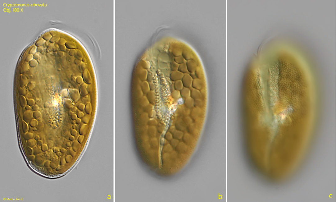
Fig. 3 a-c:Cryptomonas obovata. L = 54 µm. Three focal planes of a third specimen from left with a more obvoid shape. Obj. 100 X.
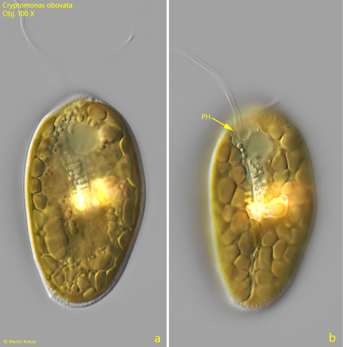
Fig. 4 a-b:Cryptomonas obovata. L = 47 µm. Two focal planes of a forth specimen from ventral. The pharynx (PH) is visible where the flagella rises. Obj. 100 X.