cell spindle-shaped, spirally twisted with 5–6 counterclockwise turns
anterior end obliquely truncated
length 42–62 µm, width 24–30 µm
leading flagellum about 80 µm long
trailing flagellum half as long as cell
periplast smooth
one contractile vacuole in anterior end
cell filled with oil droplets and rod-shaped paramylon grains
sometimes green and yellow remains of ingested algae
nucleus in the posterior end
Heteronema spirale
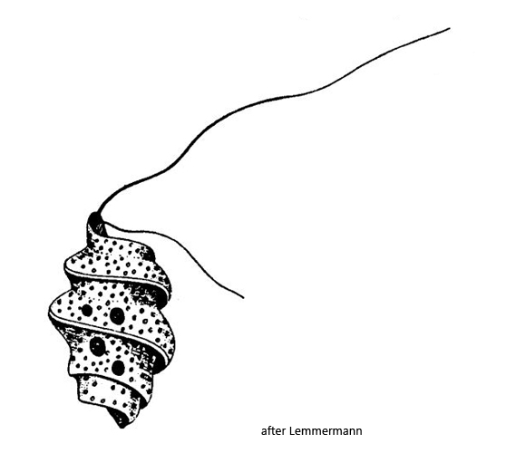
I find Heteronema spirale rarely, but regularly in the Simmelried. Mostly in samples from the upper mud layer. Because of the characteristic shape this euglenoid flagellate is easy to identify. Only Heteronema trispira has a similar spiral shape and two flagella. But this species is metabolic, has only 3 turns and is larger (96–130 µm).
According to the description of Lemmermann (1910) the spirally twisted periplast of Heteronema spirale is smooth. However, in my population I could see a distinct striation of the pellicle in all specimens I observed (s. figs. 2 a and 3 a). The lengths of the specimens in my population were between 45–65 µm and thus exactly in the range given by Lemmermann. I could also observe the 5–6, counterclockwise twists of the cell in all specimens (s. fig. 1 c). Unfortunately I could only see the long leading flagellum (s. figs. 1 a, 2 b and 3 a). Probably the trailing flagellum lies between the turns attached to the body and is therefore difficult to see.
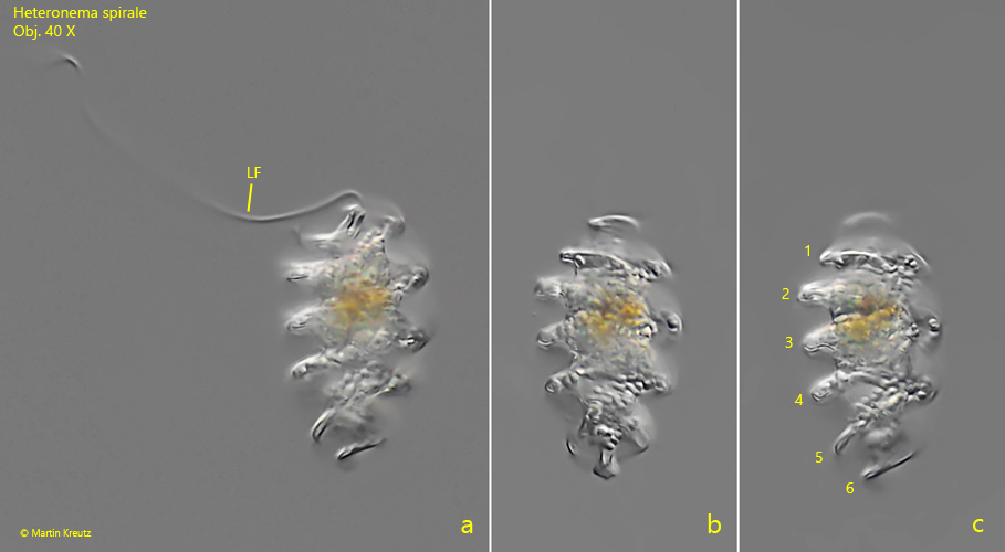
Fig. 1 a-c:Heteronema spirale. L = 47 µm. A freely swimming specimen. The cell is spirally twisted with 6 turns (1–6). LF = leading flagellum. Obj. 40 X.
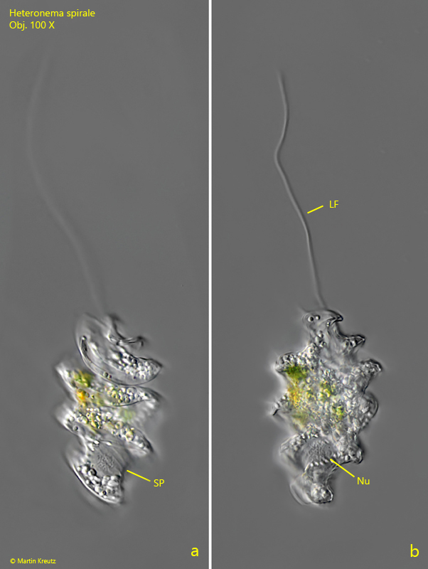
Fig. 2 a-b:Heteronema spirale. L = 55 µm. A second, slightly squashed specimen. Note the striation of the pellicle (SP). LF = leading flagellum, Nu = nucleus. Obj. 100 X.
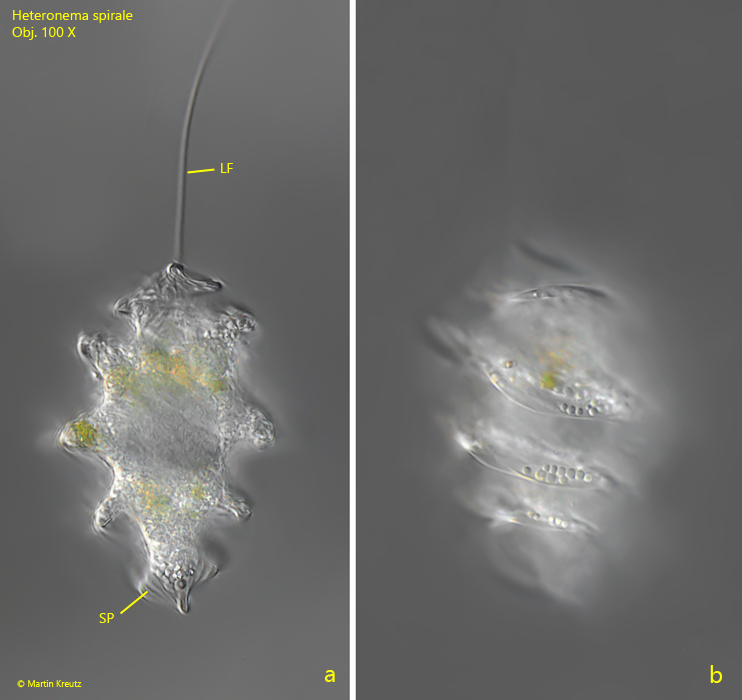
Fig. 3 a-b:Heteronema spirale. L = 63 µm. A third, freely swimming specimen. Note that the turns of the body are counterclockwise (b). LF = leading flagellum, SP = striation of the pellicle. Obj. 100 X.