In December 2023 I found Hyalogonium elongatum in a ditch overgrown with grass that was under water. The ditch was located north of the Simmelried.
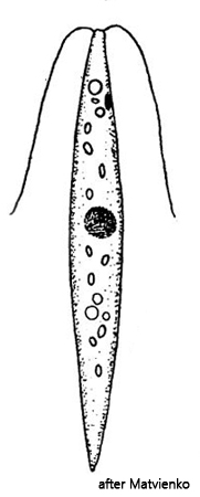
The genus Hyalogonium belongs to the volvococcal algae and is a colorless variant of the genus Chlorogonium, to which e.g. Chlorogonium elegans belongs. Within the genus Hyalogonium, 4 species have been described. The main characteristics for distinguishing these species are the presence of an eyespot, the body shape and the number and position of contractile vacuoles.
The specimens of my population are clearly slender spindle-shaped, about 50–60 µm long and have an eyespot. In addition, there are contractile vacuoles both in front of and behind the nucleus. These characteristics apply to Hyalogonium elongatum and Hyalogonium klebsii. I was able to observe at least 5 contractile vacuoles (s. figs 3 a-b and 4). Therefore, it must be Hyalogonium elongatum, as Hyalogonium klebsii has only 2 contractile vacuoles.
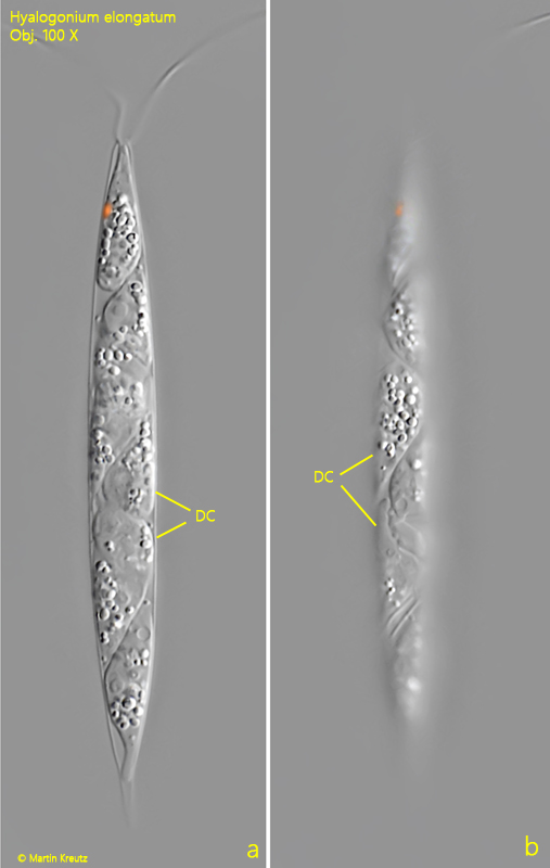
Reproduction takes place within the mother cell. After dividing three times, 8 daughter cells are formed through oblique division. As they grow, the cell wall of the mother cell finally ruptures and the daughter cells are released. I was able to observe various stages of this reproduction process (s. fig. 5 a-b).
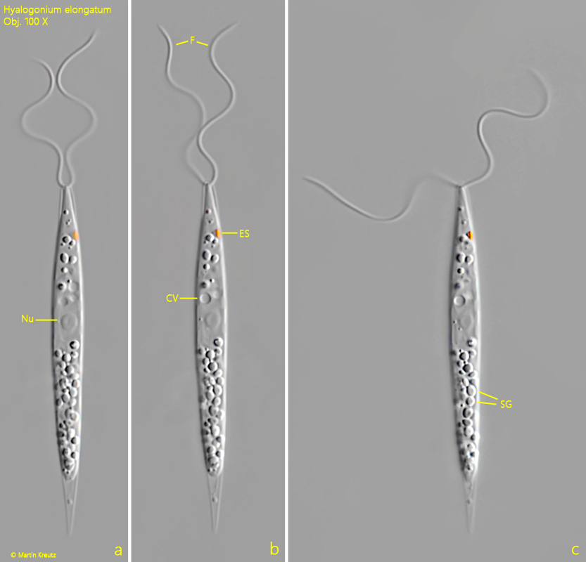
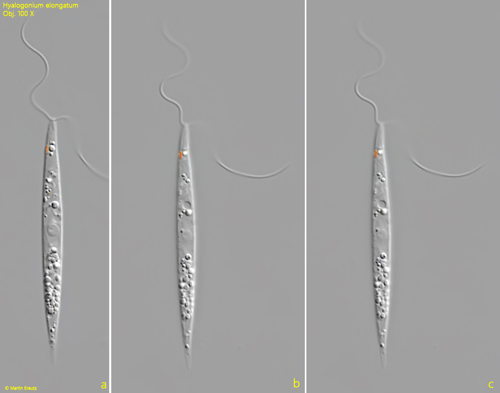
Fig. 1 a-c:Hyalogonium elongatum. L = 49 µm. A freely swimming specimen. CV = contractile vacuole, ES = eyespot, F = flagella, SG = starch grains. Obj. 100 X.
Fig. 2 a-c:Hyalogonium elongatum. L = 53 µm. A second freely swimming specimen. Obj. 100 X.
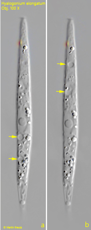
Fig. 3 a-b:Hyalogonium elongatum. L = 58 µm. The distribution of the contractile vacuoles (arrows) anteriorly to the nucleus and posteriorly. In this specimen 4 contractile vacuoles were visible. Obj. 100 X.
Fig. 4:Hyalogonium elongatum. L = 72 µm. In this specimen 5 contractile vacuoles (arrows) are visible. Obj. 100 X.
Fig. 5 a-b:Hyalogonium elongatum. L = 77 µm. Two focal planes of a specimen during the formation of daughter cells (DC). The daughter cells are released after the cell wall of the mother cell is ruptured. Obj. 100 X.