Lagynus 1
Most likely ID: Lagynus nov. spec.
Â
Synonym: n.a.
Â
Sampling location: Purren pond
Â
Phylogenetic tree: n.a.
Â
Diagnosis:Â
- body slenderly clavate
- oral bulge cone-shaped, truncated transversely
- length about 130 µm
- contractile vacuole terminal
- extrusomes clamp-shaped, about 6.5-7.5 µm long
- macronucleus kidney-shaped
- one micronucleus in concave indentation of macronucleus
- somatic cilia in widely spaced transverse rows
- cytoplasm filled with symbiotic bacteria
No drawings from previous authors available.
So far, I have only found one specimen of the haptorid ciliate shown here in November 2023 in the Purren pond. Based on the shape of the oral bulge, I believe this belongs to the genus Lagynus. The neck is not flexible like in the genus Lacrymaria. Apically, the oral bulge is truncated transversely, which is why the genera Lagynophrya, Trachelophyllum and Chaenea are not considered. Although the neck area of the ciliate is not distinctly furrowed like in Lagynus elegans, shallow, ring-shaped furrows run over the entire body. For the time being, I designate the ciliate as Lagynus 1.
Â
The body of Lagynus 1 is slender, clavate, and only slightly contractile. The cone-shaped oral bulge is approximately cylindrical in shape. The cytopharynx is lined with clamp-shaped extrusomes, which appear as two densely parallel rods (s. figs. 2 a and 3). These are also found distributed in the cytoplasm. In the cytoplasm, large quantities of symbiotic bacteria can also be found, which are especially concentrated around the area of the macronucleus (s. fig. 4 a-b). According to my measurements, they are 3–5 µm long.
Â
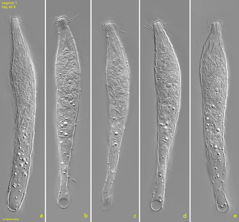
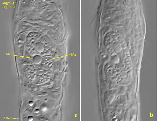
The macronucleus is kidney-shaped and is located slightly above the center of the cell. In its concave indentation lies the round micronucleus, which is noticeably highly refractive (s. fig. 5 a-b). The contractile vacuole is terminal. The somatic cilia are arranged in transverse rows. These run in shallow, ring-shaped grooves, which is why the margins of the body appear slightly constricted.
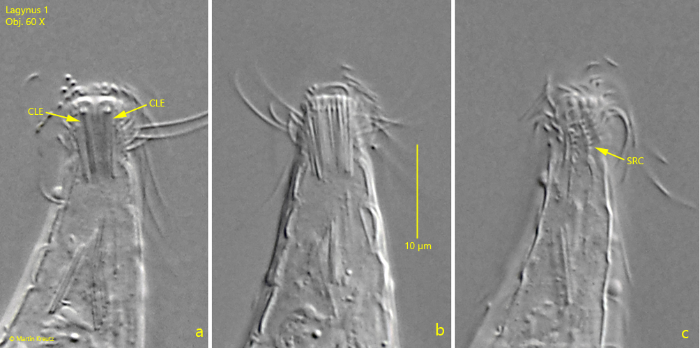
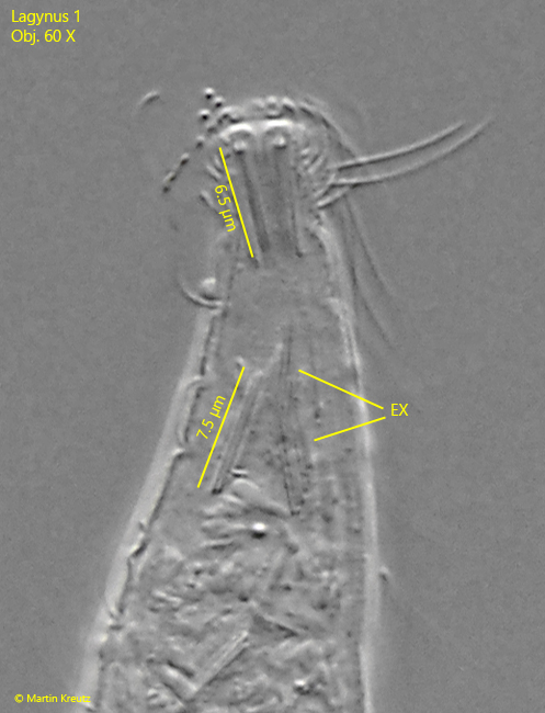
I have not been able to find a comparable species with these characteristics in the literature available to me. The genus Lagynus has been little studied so far. I therefore consider it not unlikely that this is an as yet undescribed species Lagynus nov. spec.
Fig. 1 a-e: Lagynus 1. L = 133 µm. A freely swimming specimen. Obj. 60 X.
Fig. 2 a-c: Lagynus 1. The cone-shaped oral bulge is equipped with clamp-shaped extrusomes (CLE). Slightly spiral ciliary rows (SRC) are running longitudinally from the oral bulge. Obj. 60 X.
Fig. 3: Lagynus 1. The clamp-shaped extrusomes (EX) have a length of 6.5–7.5 µm. They are also scattered in the cytoplasm. Obj. 60 X.
Fig. 4 a-b: Lagynus 1. The cytoplasm is filled with rod-shaped symbiotic bacteria (SB) with a length of 3–4 µm. The symbiotic bacteria are concentrated in the area of the macronucleus. Obj. 60 X.
Fig. 5 a-b: Lagynus 1. The macronucleus (Ma) is kidney-shaped. In the concave indentation of the macronucleus the highly refractive micronucleus (Mi) is located. Obj. 60 X.