ciliation uniform, 80–120 ciliary rows of paired cilia
a tuft of caudal cilia
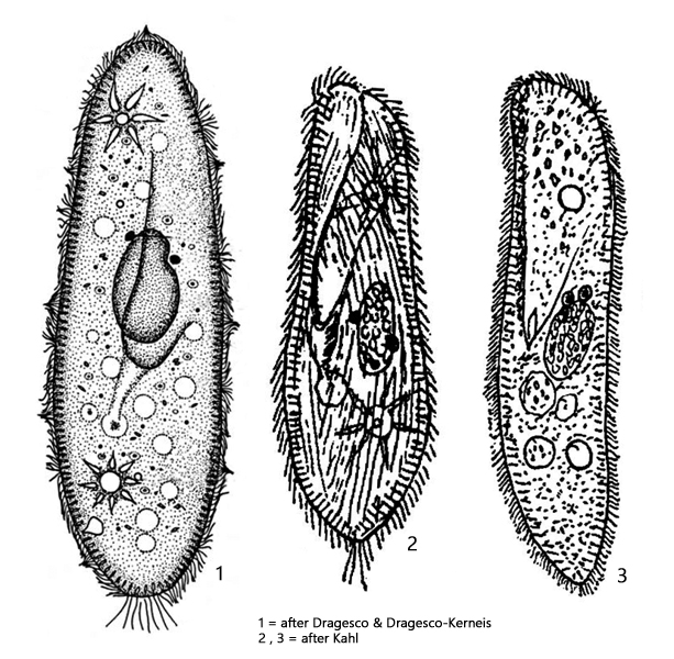
Paramecium aurelia
I found Paramecium aurelia in an old sample containing decomposing water lily leaves. This finding corresponds with Kahl’s (1935) description that Paramecium aurelia often occurs in samples containing decaying plants.
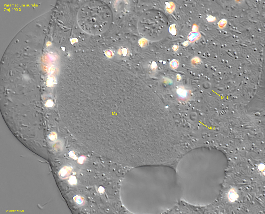
Paramecium aurelia is slightly smaller than Paramecium caudatum and is a fast, restless swimmer. Its characteristics are essentially the same as those of Paramecium caudatum, which is why the species has certainly been overlooked frequently. The only reliable identifying feature is characterstic of the the nuclear apparatus. There are a large, ellipsoid macronucleus and two micronuclei, each are located in separate vacuoles. However, this can only be seen in squashed specimens (s. figs. 3 and 4).
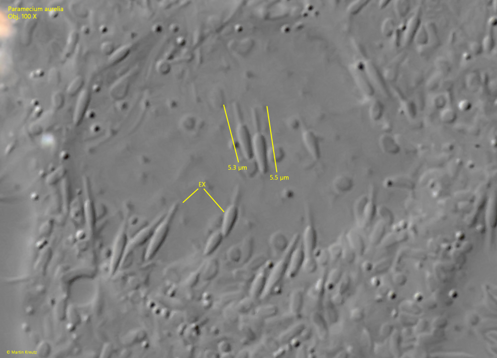
The specimens in my population were on average 150 µm long. At 5.3–5.5 µm, the extrusomes were slightly longer than those described by Foissner, Berger & Kohmann (1994), who give a length of 3–4 µm (s. fig. 7). Otherwise, all other characteristics were consistent.
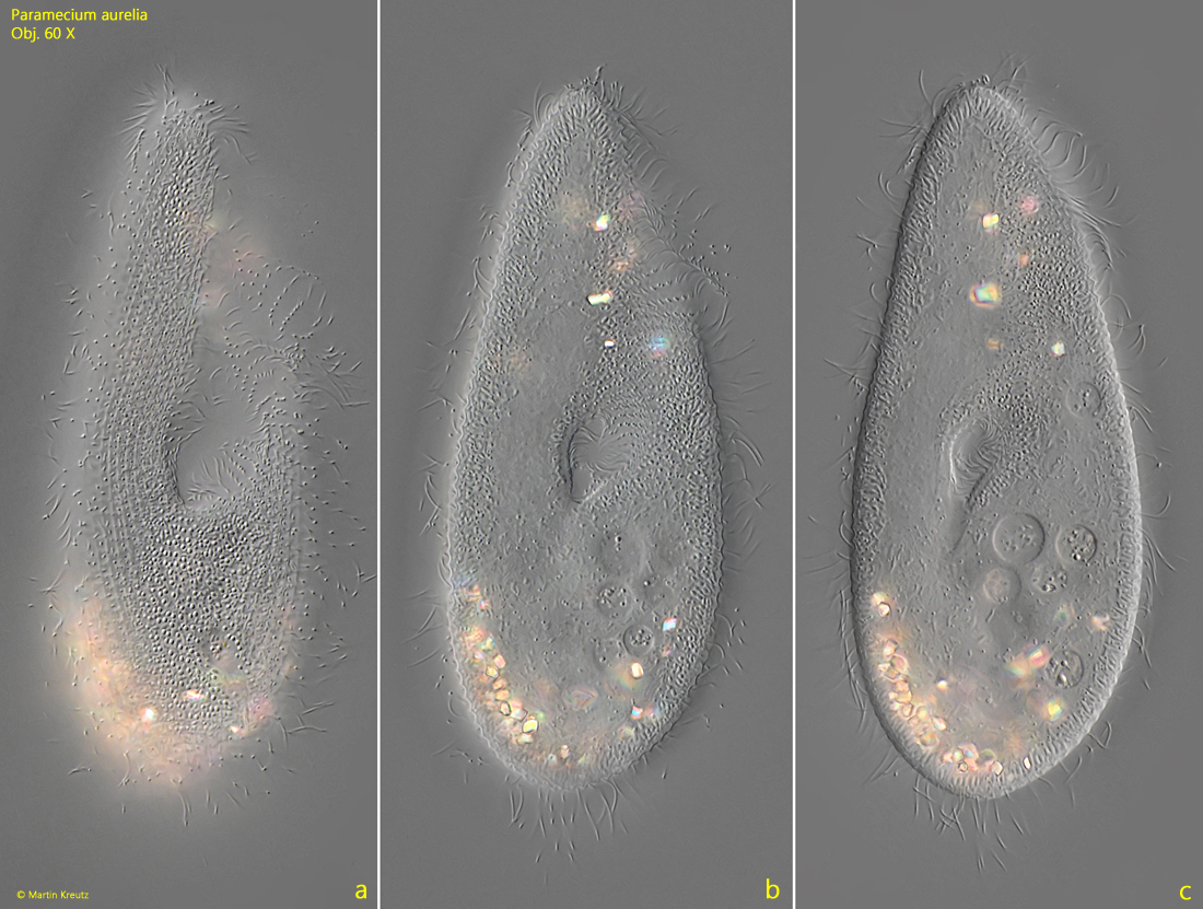
Fig. 1 a-c:Paramecium aurelia. L = 155 µm. Three focal planes of a freely swimming specimen from ventral. Obj. 60 X.
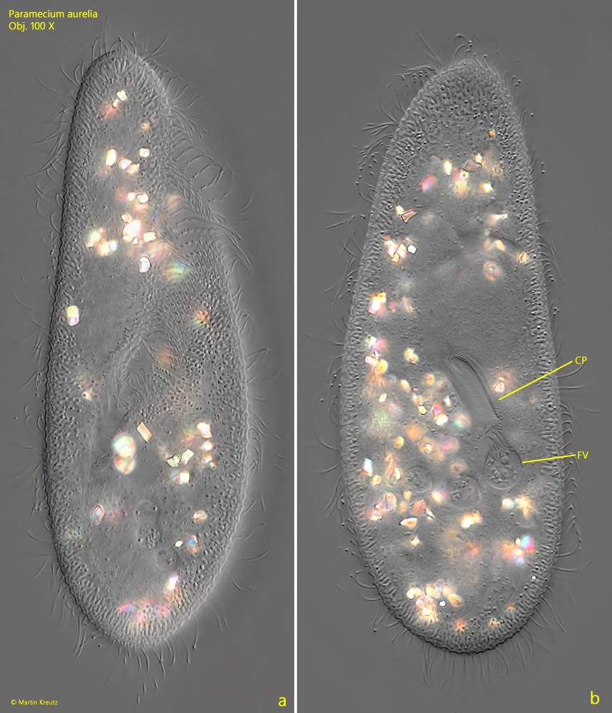
Fig. 2 a-b:Paramecium aurelia. L = 156 µm. A second freely swimming specimen from ventral (a) and from dorsal (b). The cytopharynx (CP) fills a food vacuole (FV) with collected bacteria. Obj. 100 X.
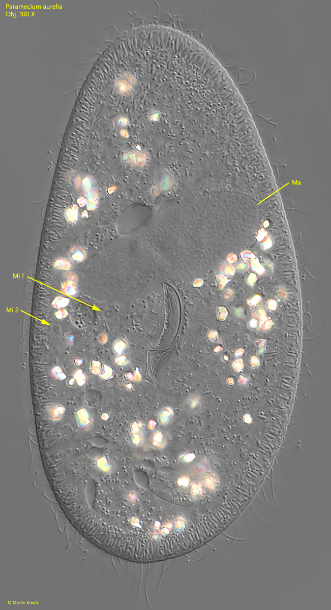
Fig. 3:Paramecium aurelia. The ellipsoid macronucleus (Ma) and the two micronuclei (Mi 1, Mi 2) in a second squashed specimen. Obj. 100 X.
Fig. 4:Paramecium aurelia. The ellipsoid macronucleus (Ma) and the two micronuclei (Mi 1, Mi 2) in a squashed specimen. The miconuclei are enclosed in separate vacuoles. Obj. 100 X.
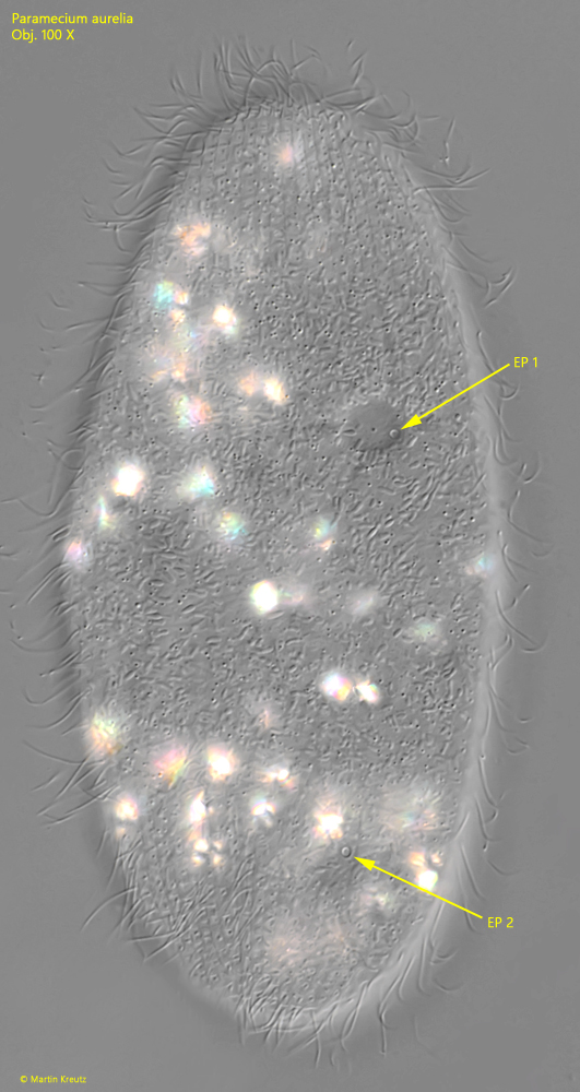
Fig. 5:Paramecium aurelia. The two excretion pores (EP 1, EP 2) of the two contractile vacuoles are located dorsally. Obj. 100 X.
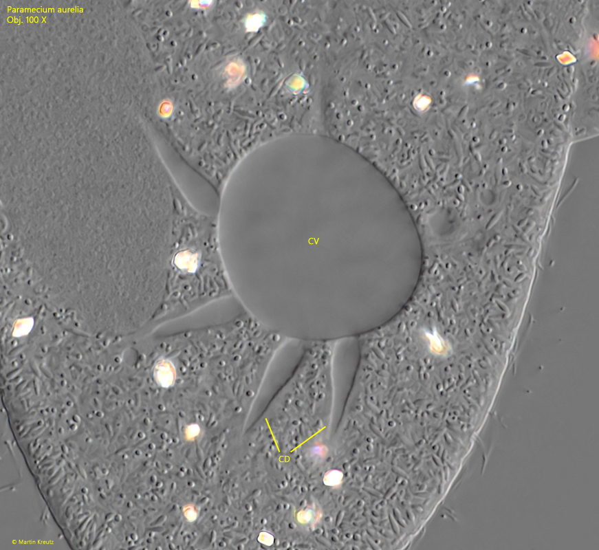
Fig. 6:Paramecium aurelia. The posterior contractile vacuole (CV) in a squashed specimen with several collection ducts (CD). Obj. 100 X.
Fig. 7:Paramecium aurelia. The extrusomes (EX) in a squashed specimen are 5.3–5.5 µm long. Obj. 100 X.
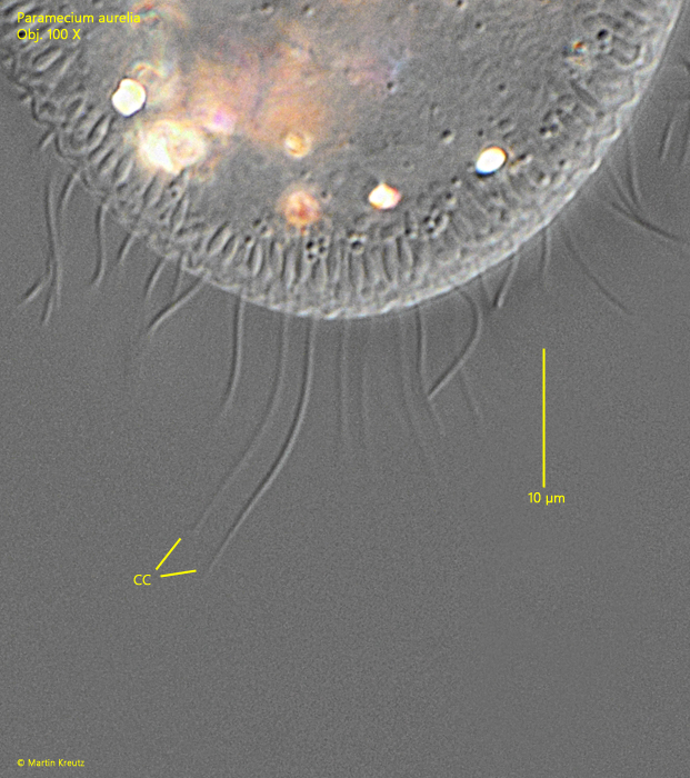
Fig. 8:Paramecium aurelia. The caudal cilia with a length of 15–17 µm. Obj. 100 X.
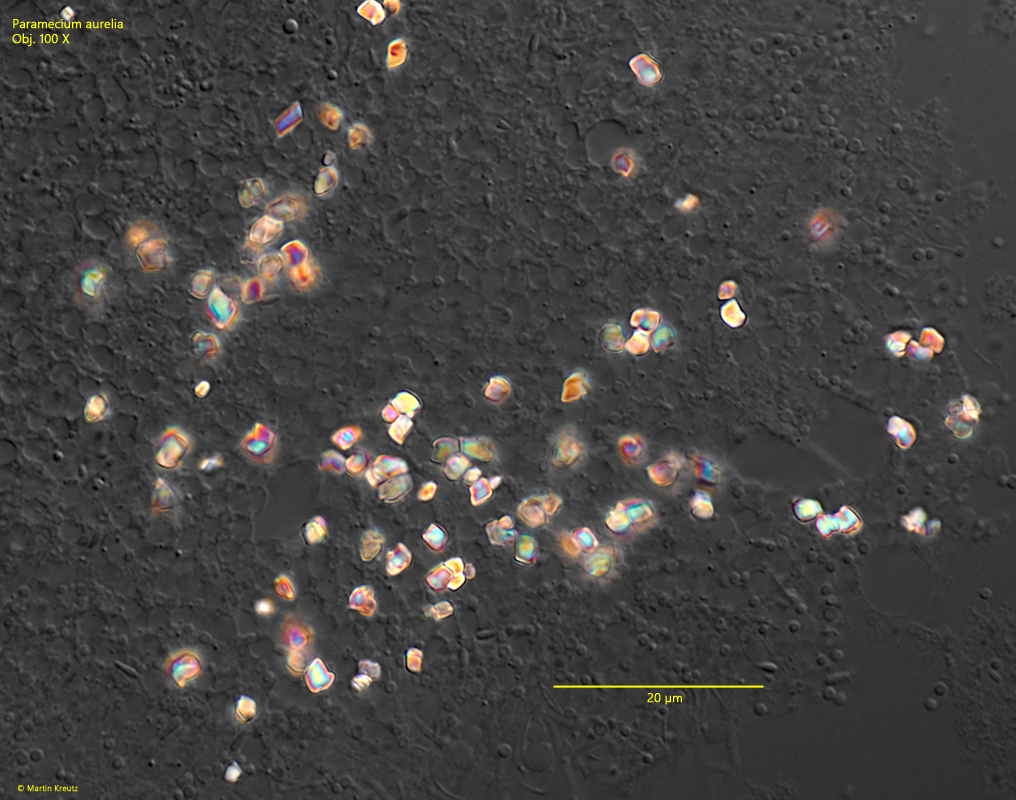
Fig. 9:Paramecium aurelia. The crystals scattered in the cytoplasm have a diameter of about 2–4 µm and a more or less rectangular shape. Obj. 100 X.