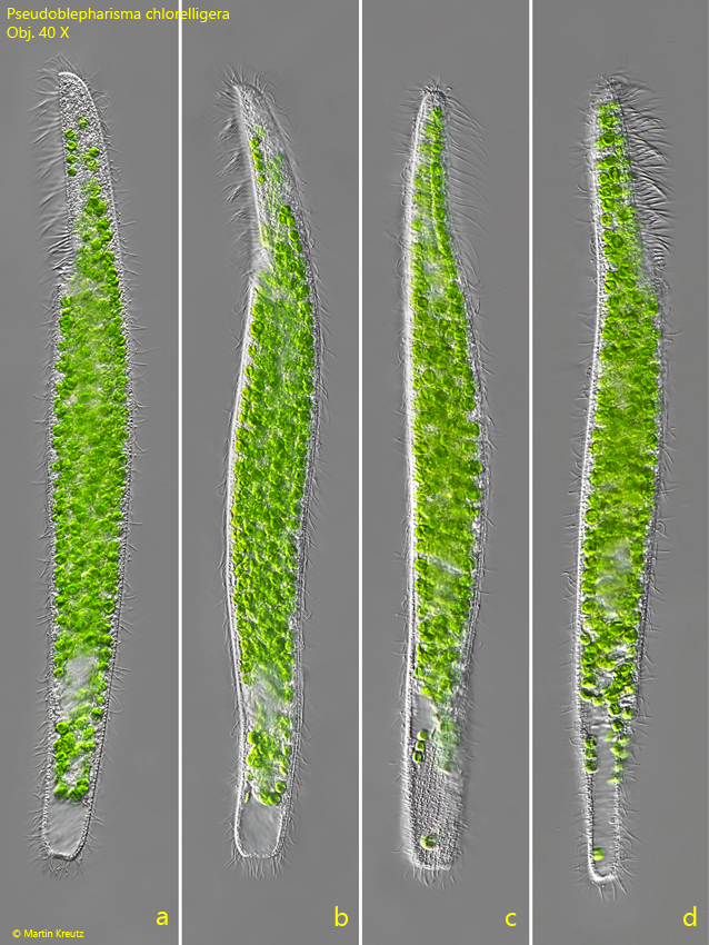
Pseudoblepharisma chlorelligera

(Sramek-Husek, 1949) Muñoz-Gómez et al., 2025

Most likely ID: n.a.


Synonyms: Pseudoblepharisma tenue var. chlorelligera, Pseudoblepharisma tenue var. viride


Sampling location: Simmelried


Phylogenetic tree: Heterotrichida


Diagnosis: 

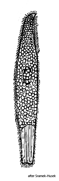
  • body elongated, parallel-sided, posterior end bluntly rounded
  • body 50% contractile
  • length 195–362 µm, width 28–36 µm
  • adoral zone reach one fourth of body length
  • to right side of adoral zone a undulating membrane of short cilia
  • cytoplasm green due to symbiotic algae
  • numerous symbiotic bacteria type 1 scattered in cytoplasm
  • numerous symbiotic bacteria type 2 in alveoli beneath pellicle
  • 10-12 longitudinal rows of cilia
  • between the rows of cilia stripes of refractive granules
  • macronucleus ellipsoid, near mid-body
  • several micronuclei adjacent to the macronucleus
  • contractile vacuole large, terminal, with canal
Pseudoblepharisma-chlorelligera
Pseudoblepharisma chlorelligera
I find Pseudoblepharisma chlorelligera exclusively in my collection area Simmelried, where this species is very common. I have been able to continuously detect Pseudoblepharisma chlorelligera there since 1993, with strong fluctuations in population density. Between 2005 and 2010 there was a strong maximum. Since then, the population density has been continuously declining. The specimens are most often found in the uppermost layer of mud and in the underlying, oxygen-poor zones.

Pseudoblepharisma chlorelligera was first described by Sramek-Husek in 1949 as Pseudopblepharisma tenue var. chlorelligera. His description, written in Czech, is extremely brief. Previously, in 1926, the species Pseudoblepharima tenue was described by Kahl. This similar species is distinctly pink-colored due to numerous symbiotic pink-colored bacteria. The variant described by Sramek-Husek, on the other hand, is intensely green-colored due to numerous symbiotic algae in the cytoplasm. Based on microscopic and genetic analyses by Munoz-Gomez et al. (2025), it was elevated to the species Pseudoblepharisma chlorelligera.
Tab. 1: Dimensions of Pseudoblepharisma tenue and number of symbiotic algae per cell.

Length (µm)

Width (µm)

No. symbiotic algae

280

29

328

310

32

401

240

32

310

334

30

482

195

35

330

254

32

266

236

36


245

29


362

32


220

34


215

34


234

28


301

30


223

35


242

29


x̄ = 259 µm

x̄ = 32 µm

x̄ = 352


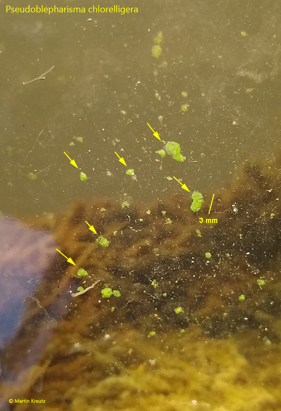
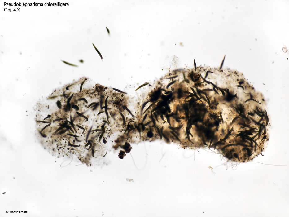
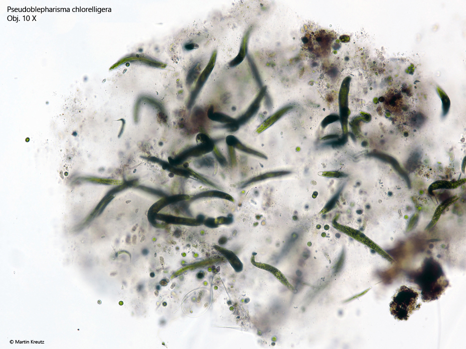
In samples with very dense populations, I was able to find the formation of mucilaginous clusters on the walls of the sample vessel, which were densely inhabited by Pseudoblepharisma chlorelligera (s. figs 1, 2 and 3). These clusters were preferentially formed on the side of the vessel facing the light. They were on average 1–3 mm in size and formed by the ciliate itself and were not subsequently colonized. This is related to the ability of Pseudoblepharisma chlorelligera to form a gelatinous lorica (s. below).


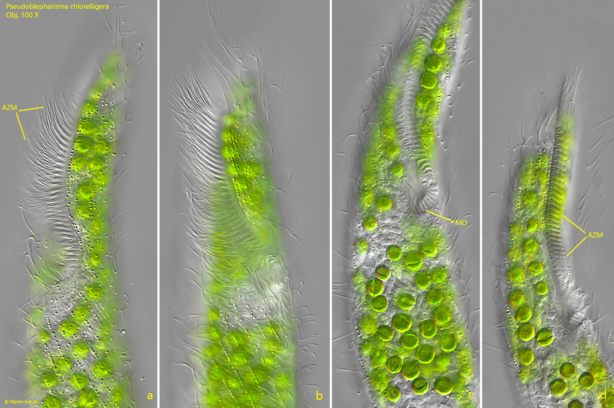
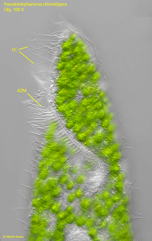
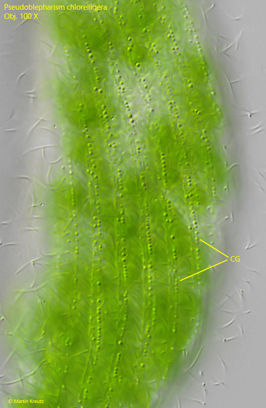
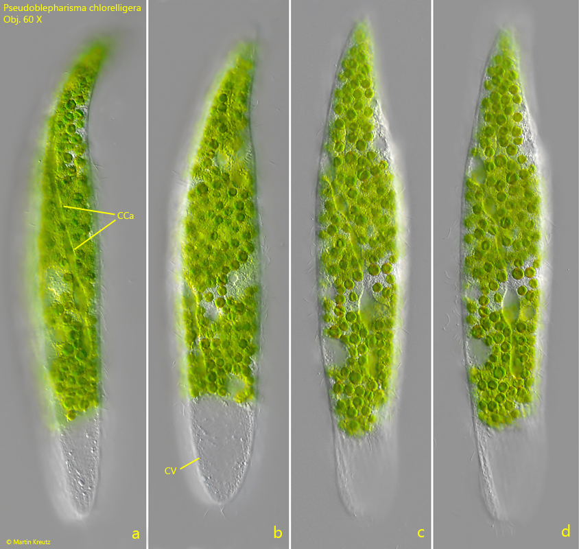
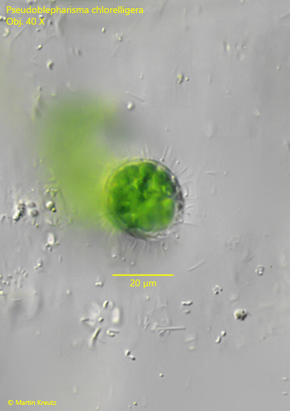
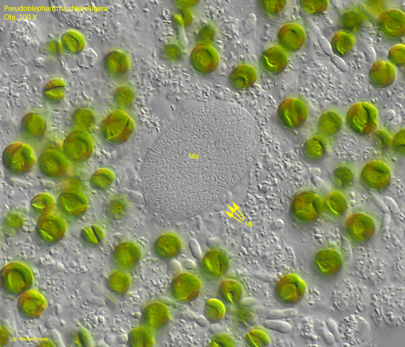
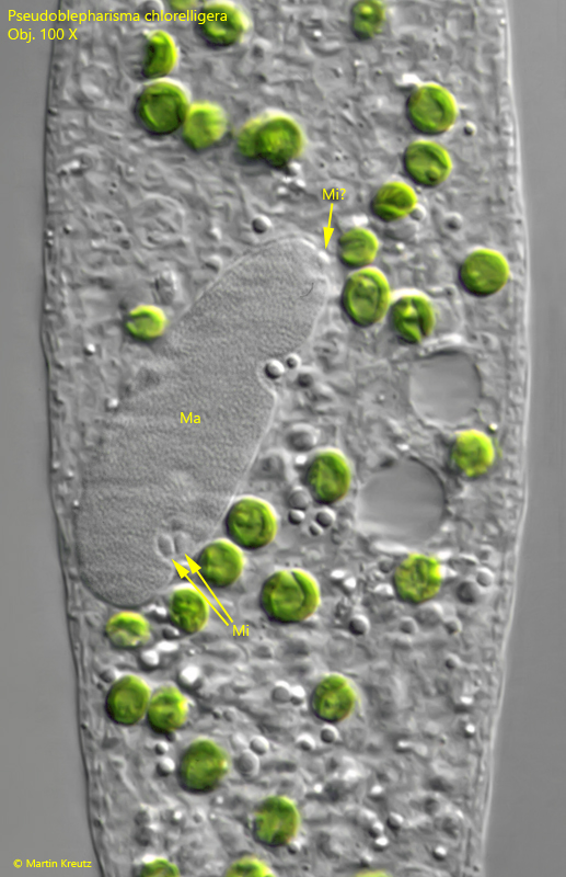
Since the description by Sramek-Husek is so brief, I have measured 15 specimens of Pseudoblepharisma chlorelligera myself (s. tabl. 1, below). The specimens of my population were 195–362 µm long, which fits well with the range of 200–300 µm given by Sramek-Husek. The width of my specimens was 28–36 µm. The ciliate is rotund and not flattened (s. fig. 10). Only the area of the terminal contractile vacuole flattens during emptying. In addition, the contractile vacuole has a collecting canal that runs on the dorsal side and extends almost to the anterior end (s. fig. 9 a-d). The adoral membranelle zone runs in a shallow groove and reaches to the front quarter of the body (s. fig. 6 a-d). There it then bends to the right toward the mouth opening. The undulating membrane on the left side of the groove is difficult to discern. In addition, on the left side of the mouth groove there is a row of strongly elongated cilia (s. fig. 7). These could possibly be sensory or tactile cilia. According to my observations, the pellicle has about 16–20 rows of cilia, between which rows of granules are located, which are colorless or slightly yellowish (s. fig. 8). The rows of cilia run slightly spirally counterclockwise around the body. The macronucleus is mostly oval or ellipsoid and about 28 µm long and 10 µm wide. It is always accompanied by some round or lens-shaped micronuclei, which are either attached to it or lie in a cavity embedded in the macronucleus (s. figs. 11 and 12).

Pseudoblepharisma-chlorelligera

Fig. 1: Pseudoblepharisma chlorelligera. Macroscopic mucilaginous clusters of cells (arrows) growing on the glass wall of the sample container. 
Pseudoblepharisma-chlorelligera

Fig. 2: Pseudoblepharisma chlorelligera. One of the mucilaginous clusters as shown in fig. 1 populated with about 100 specimens. Obj. 4 X.  
Pseudoblepharisma-chlorelligera

Fig. 3: Pseudoblepharisma chlorelligera. One of the mucilaginous clusters as shown in fig. 2 in detail. Obj. 10 X.  
Pseudoblepharisma-chlorelligera

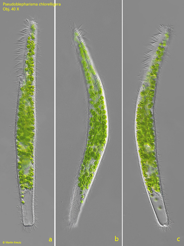
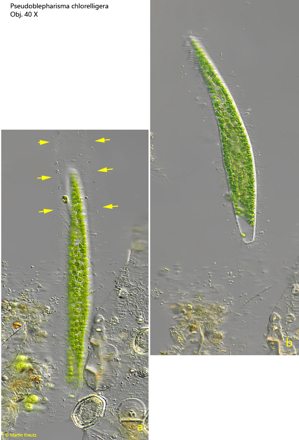
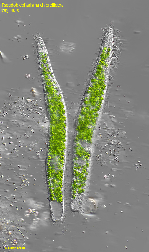
Fig. 4 a-c: Pseudoblepharisma chlorelligera. L = 307 µm. A freely swimming specimen. Obj. 40 X.  
Pseudoblepharisma-chlorelligera

Fig. 5 a-d: Pseudoblepharisma chlorelligera. L = 338 µm. A second freely swimming specimen. Obj. 40 X.  
Pseudoblepharisma-chlorelligera

Fig. 6 a-d: Pseudoblepharisma chlorelligera. Different views of the oral apparatus with the adoral zone of membranelles (AZM) and the mouth opening (MO). Obj. 100 X.  
Pseudoblepharisma-chlorelligera

Fig. 7: Pseudoblepharisma chlorelligera. On the left side of the adoral zone of membranelles (AZM) a row of elongated cilia (EC) is located. Probably they have a sensory or tactile function. Obj. 100 X.  
Pseudoblepharisma-chlorelligera

Fig. 8: Pseudoblepharisma chlorelligera. The cortical granules (CG) are colorless and arranged in stripes between the longitudinal ciliary rows. Obj. 100 X.  
Pseudoblepharisma-chlorelligera

Fig. 9 a-d: Pseudoblepharisma chlorelligera. The collecting canal (CCa) of the contractile vacuole (CV) is located dorsally and reach almost the anterior end. Obj. 40 X.  
Pseudoblepharisma-chlorelligera

Fig. 10: Pseudoblepharisma chlorelligera. In the optical cross-section of the body, it can be seen that it is rotationally symmetrical and not flattened. Obj. 40 X.  
Pseudoblepharisma-chlorelligera

Fig. 11: Pseudoblepharisma chlorelligera. The ellipsoid macronucleus (Ma) with several adjacent micronulcei (Mi). Obj. 100 X.
Pseudoblepharisma-chlorelligera

Fig. 12: Pseudoblepharisma chlorelligera. The macronucleus (Ma) of a second specimen with at least 2 adjacent micronuclei (Mi). Mi? = probably a third micronucleus. Obj. 100 X.
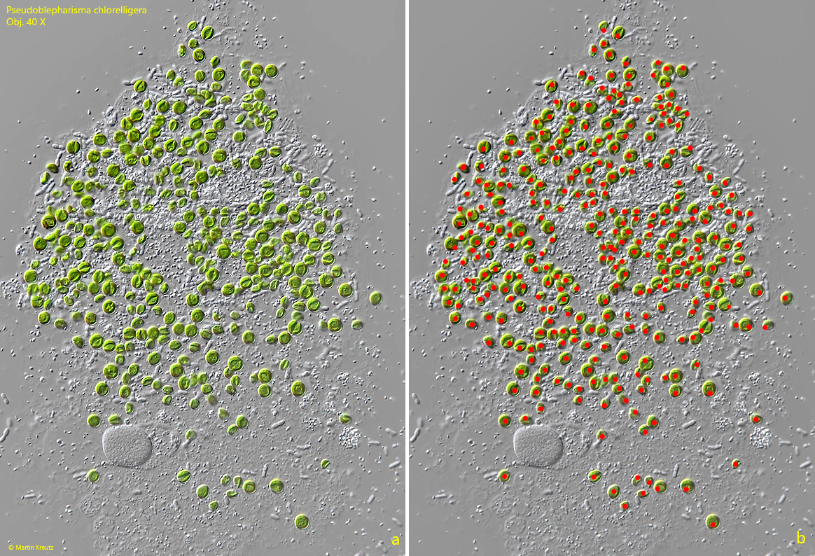
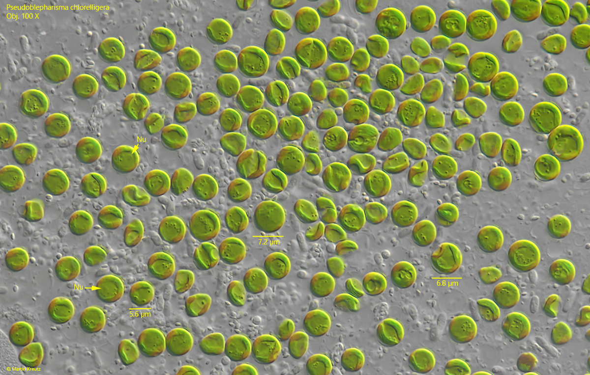
The most striking feature of Pseudoblepharisma chlorelligera are the symbiotic algae, which color the ciliate a vivid green. At first glance, one might think there are about 50–100 algal cells. However, if a specimen is squashed strongly and count them, there are significantly more (s. fig. 13). I counted the number of symbiotic algae in 6 specimens (s. tabl 1, above). They number range between 266–482 algae cells per specimen (x = 352). The algae have a diameter of about 5-7.5 µm and belong to the genus Chlorella. They possess their own nucleus (s. fig. 14).

Pseudoblepharisma-chlorelligera

Fig. 13: Pseudoblepharisma chlorelligera. To count the symbiotic algae, the specimen was heavily crushed to separate the algae. To avoid double counting during the counting process, each counted alga was marked with a red dot. This specimen had 328 symbiotic algae. Obj. 40 X.
Pseudoblepharisma-chlorelligera

Fig. 14: Pseudoblepharisma chlorelligera. The symbiotic algae in a strongly squashed specimen are from the Chlorella type with an own nucleus (Nu). The algae cells have a diameter of 5.6–7.2 µm. Obj. 100 X.
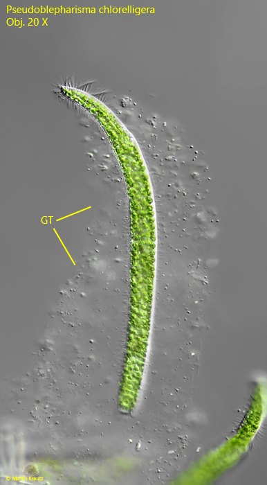
In fresh samples, freely swimming specimens of Pseudoblepharisma chlorelligera are usually found. However, if a sample is placed in a petri dish and floating coverslips are laid on it, after a few days it becomes colonized and one can observe how Pseudoblepharisma chlorelligera builds its own gelatinous lorica. This lorica is usually tubular, but sometimes conical (s. figs. 15, 16 a-b and 17). In the similar species Pseudoblepharisma tenue, no construction of a lorica has been observed so far.

Pseudoblepharisma-chlorelligera

Fig. 15: Pseudoblepharisma chlorelligera. L =  340 µm. A specimen in a self contructed gelatinous tube (GT). Obj. 20 X. 
Pseudoblepharisma-chlorelligera

Fig. 16 a-b: Pseudoblepharisma chlorelligera. L = 226 µm. A second specimen is moving in the self contructed, tube-shaped lorica. The delicate wall of the tube is only visible by the attached bacteria and granules (arrows). Obj. 40 X. 
Pseudoblepharisma-chlorelligera

Fig. 17 a-b: Pseudoblepharisma chlorelligera. L = 288 µm + 304 µm. Two specimens in their gelatinous tubes. Obj. 40 X. 
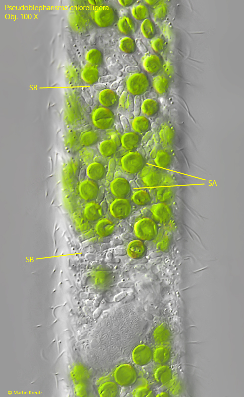
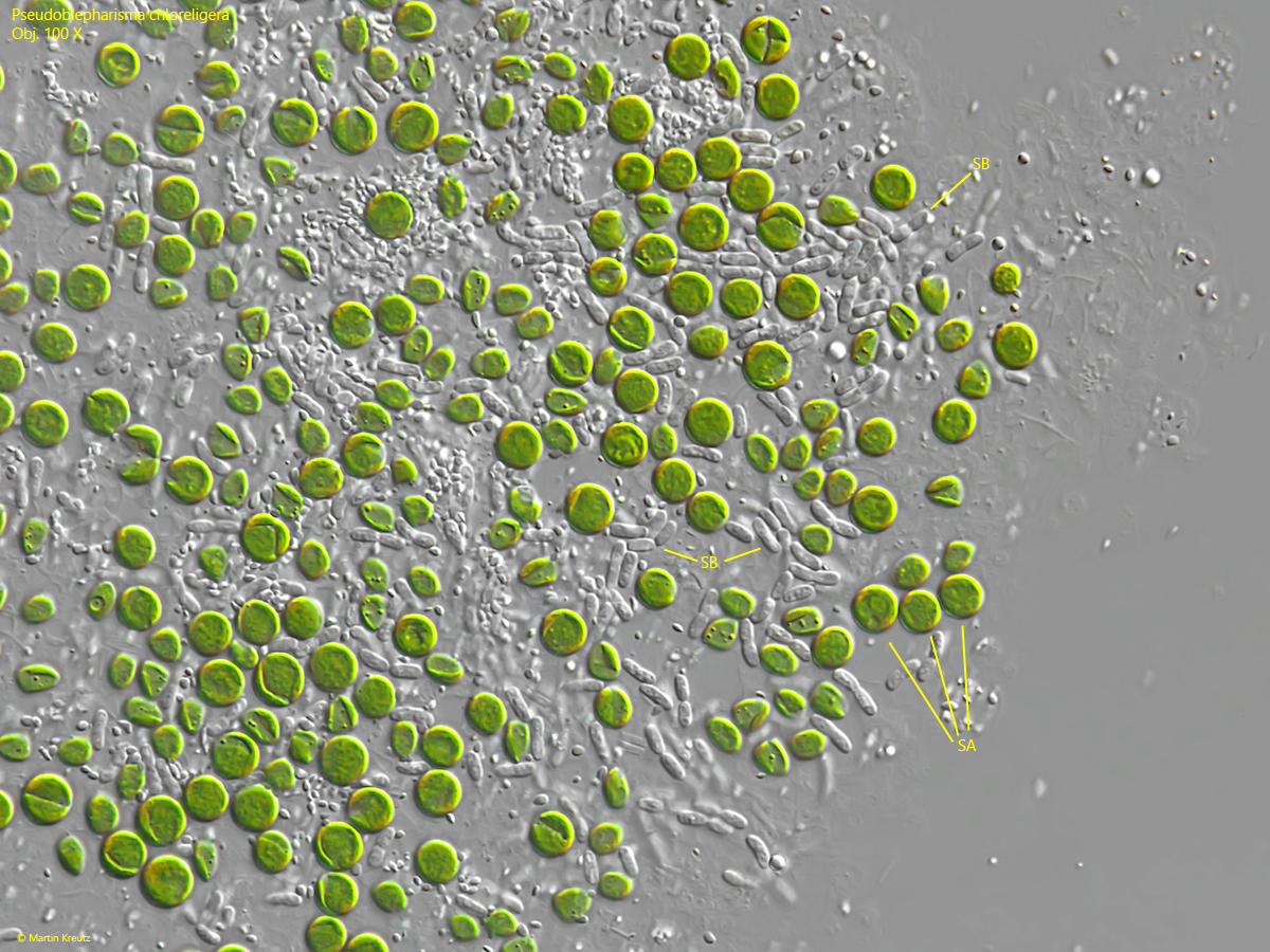
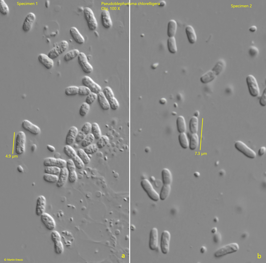
At higher magnification, it can be seen that there are also quite large bacteria present in the cytoplasm of Pseudoblepharisma chlorelligera, which, like the symbiotic algae, are distributed throughout the cytoplasm (s. fig. 18). These are somewhat irregularly shaped rods with rounded ends and a length of 3–5 µm (s. figs. 19 and 20 a-b).


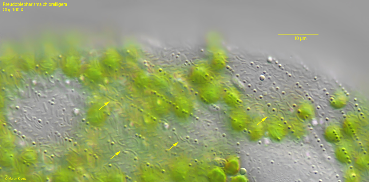
The presence of two symbionts is very rare in ciliates. Therefore, Pseudoblepharisma chlorelligera was thoroughly examined microscopically and genetically by Muñoz-Gómez et al. (2025) to learn more about the relationship between the ciliate and its symbionts as well as between the symbionts themselves. However, a gene analysis revealed that not only one prokaryotic genome was present, but even two. This indicated a second, previously undiscovered prokaryotic symbiont in Pseudoblepharisma chlorelligera. This second bacterium was then discovered in the alveoli of the ciliate, just beneath the pellicle, using DAPI (4′,6-diamidino-2-phenylindole) staining. This second prokaryotic symbiont consists of slightly curved, thin rods, about 2.5 µm in length and about 0.4 µm in thickness. These curved bacteria can also be recognized without staining at high magnification (s. fig. 21). The bacteria distributed in the cytoplasm were initially designated as candidatus Accumulibacter symbioticus, and the bacteria in the alveoli as candidatus Propionivibrio subcutaneus. This would make Pseudoblepharisma chlorelligera one of the very rare cases of a ciliate with three symbionts.
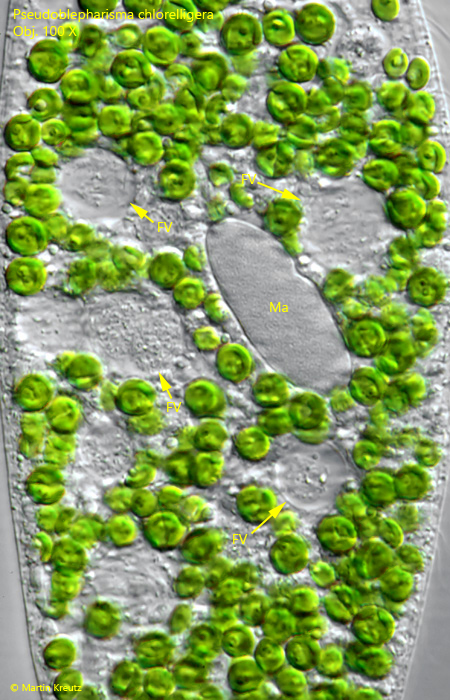
 

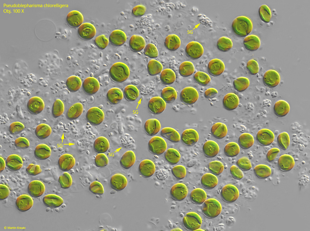
Muñoz-Gómez et al. (2025) also attempted to decipher the complex metabolic relationship between the host and its symbionts. Candidatus Accumulibacter symbioticus is a polyphosphate-accumulating bacterium, which is stored as PHB (poly-β-hydroxybutyrate) in the form of granules in the cytoplasm. The PHB is polymerized from acetate and serves as a carbon storage. These PHB granules can be recognized under a light microscope. Their concentration in the bacteria varies depending on the degree of accumulation (s. fig. 20 a-b). The second symbiotic bacterium, candidatus Propionivibrio subcutaneus, is capable of nitrogen and sulfur metabolism and is probably a supplier of essential amino acids for the host. The symbiotic algae were finally identified as Chlorella (type K10) and provide sugars and other carbohydrates through photosynthesis, which are stored by Pseudoblepharisma chlorelligera as starch grains found distributed throughout the cytoplasm (s. fig. 22). Through this combination, Pseudoblepharisma chlorelligera is supplied with both carbohydrates and amino acids. Nevertheless, this ciliate is mixotrophic, as food vacuoles filled with phagocytosed bacteria are also found in the cytoplasm (s. fig. 23).

Pseudoblepharisma-chlorelligera

Fig. 18: Pseudoblepharisma chlorelligera. In a slightly squashed specimen the symbiotic bacteria type 1 (SB, = candidatus Accumulibacter symbioticus) become visible between the symbiotic algae (SA). Obj. 100 X. 
Pseudoblepharisma-chlorelligera

Fig. 19: Pseudoblepharisma chlorelligera. In a strongly squashed specimen the symbiotic algae (SA) as well as the symbiotic bacteria type 1 (SB, = candidatus Accumulibacter symbioticus) scattered throughout the cytoplasm are visible. Obj. 100 X. 
Pseudoblepharisma-chlorelligera

Fig. 20 a-b: Pseudoblepharisma chlorelligera. The symbiotic bacteria type 1 (= candidatus Accumulibacter symbioticus) of two differents specimens. The bacteria have a different content of PHB granules ( (poly-β-hydroxybutyrate). Obj. 100 X. 

Pseudoblepharisma-chlorelligera

Fig. 21: Pseudoblepharisma chlorelligera. The symbiotic bacteria type 2 (arrows, = candidatus Propionivibrio subcutaneus) are curved rods and located in the alveoli beneath the pellicle. Obj. 100 X.

Pseudoblepharisma-chlorelligera

Fig. 22: Pseudoblepharisma chlorelligera. The carbon hydrates provided by the symbiotic algae are stored as starch in the cytoplasm. Clusters of small starch grains (SG) are visible between the symbiotic algae. Obj. 100 X.

Pseudoblepharisma-chlorelligera

Fig. 23: Pseudoblepharisma chlorelligera. Between the symbiotic algae food vacuoles (FV) are located filled with ingested bacteria. Ma = macronucleus. Obj. 100 X.

Pseudoblepharisma-chlorelligera

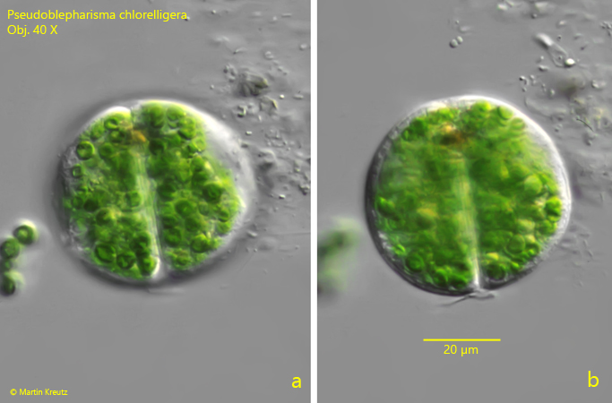
Fig. 24 a-b: Pseudoblepharisma chlorelligera. Two different focal planes of a division cyst with a diameter of 55 µm. Obj. 100 X.

Pseudoblepharisma chlorelligera

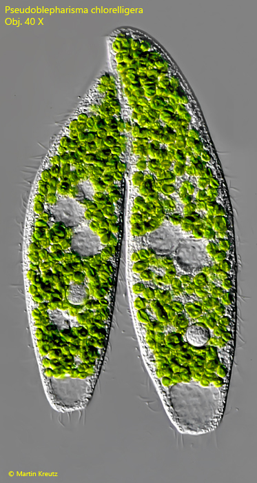
Fig. 25: Pseudoblepharisma chlorelligera. L = 175 + 192 µm. Two specimens in the process of conjugation. Obj. 40 X.