cells about 50 µm in diameter (without sheat of scales)
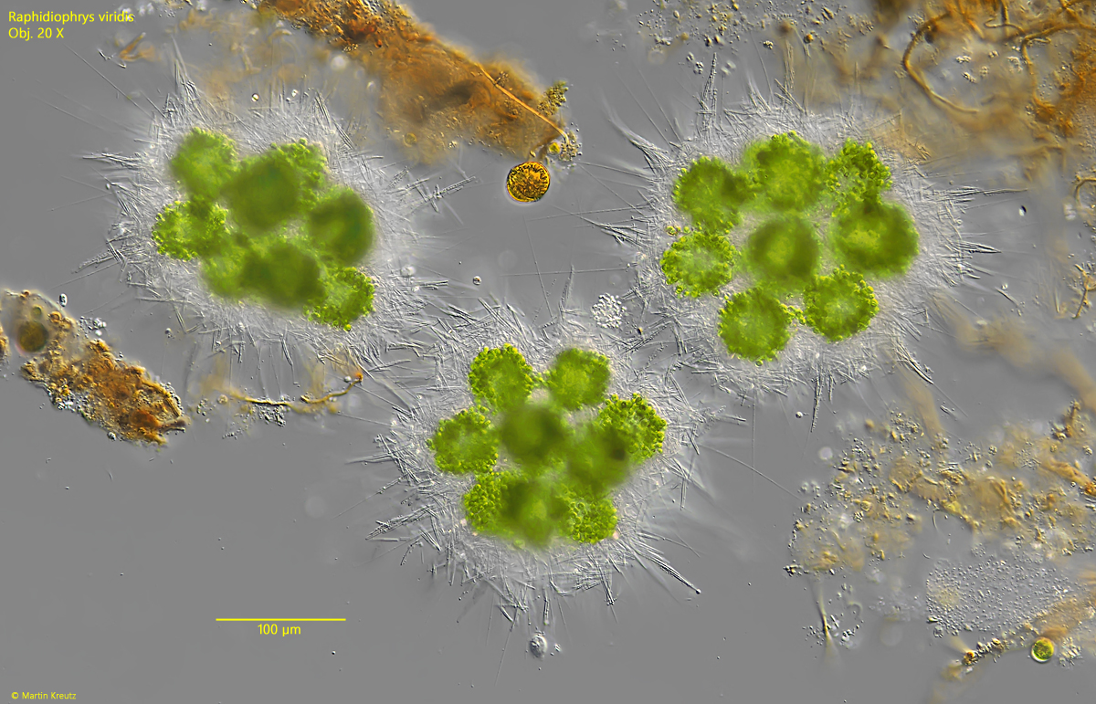
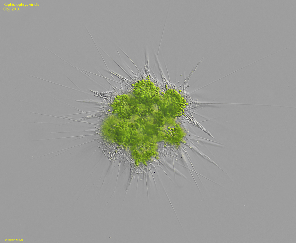
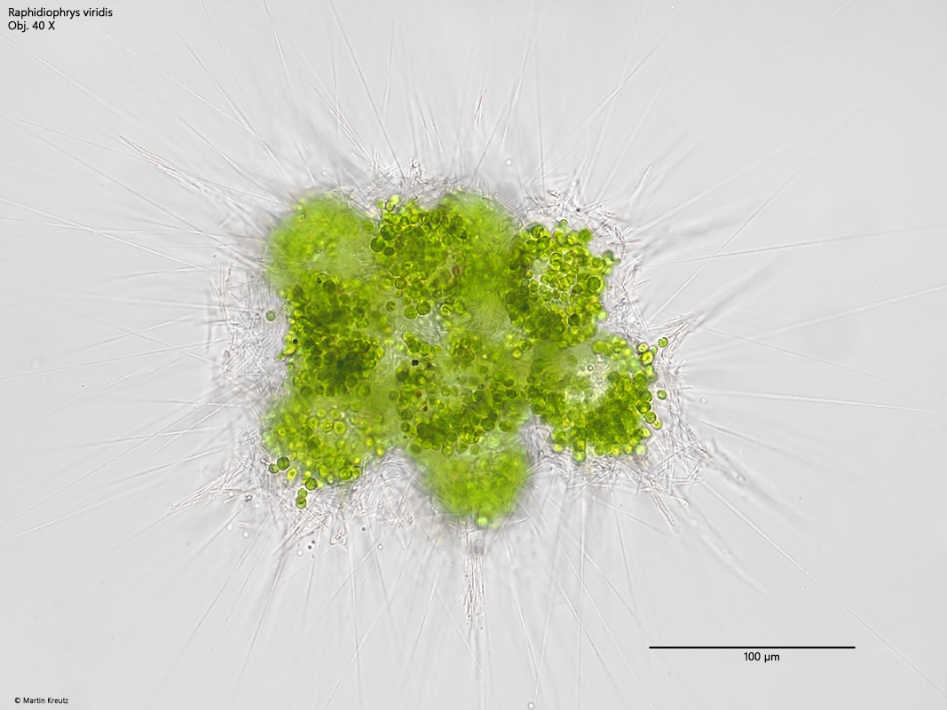
colonies of 5–18 specimen, tightly packed
diameter of colonies about 200 µm
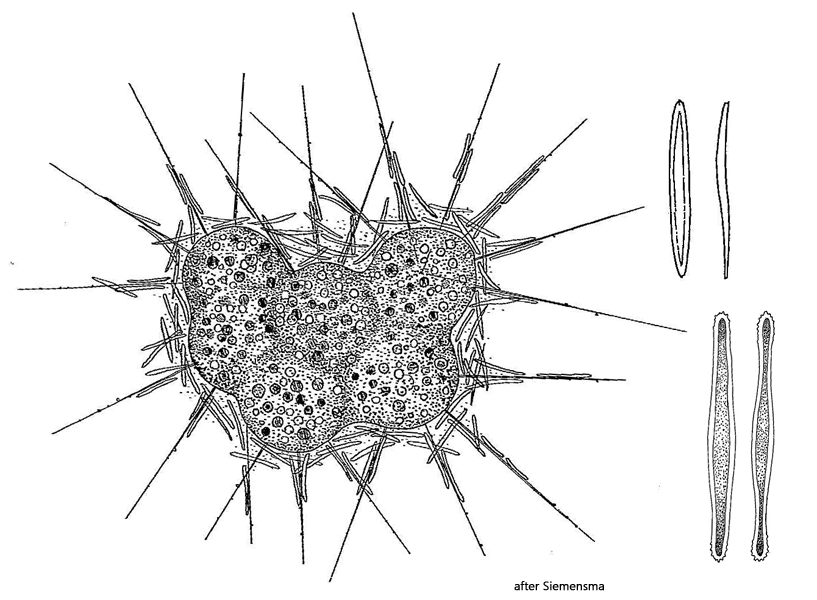
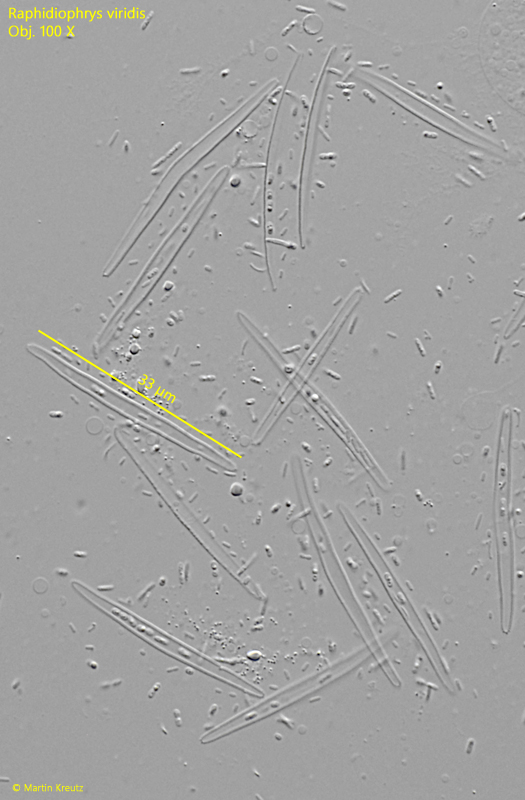
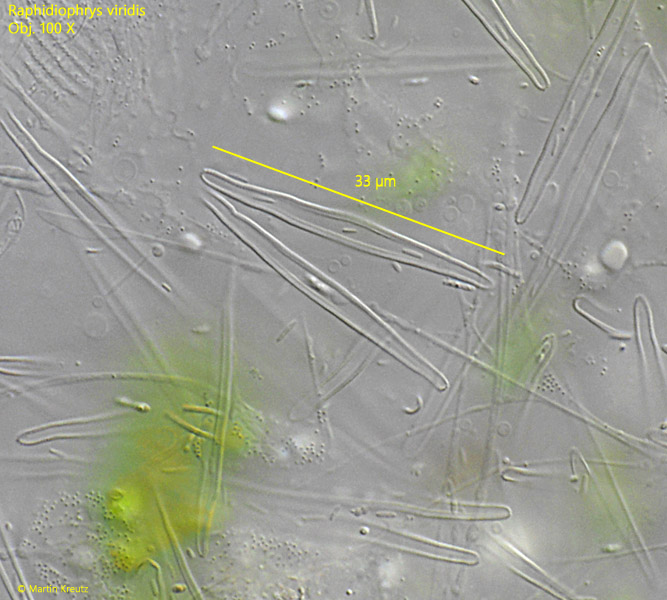
scales rod-like, with slightly tapered ends
scales 15–37 µm long, width about 3 µm
poles of scales with small papillae (hard to see)
cytoplasm green due to symbiotic algae
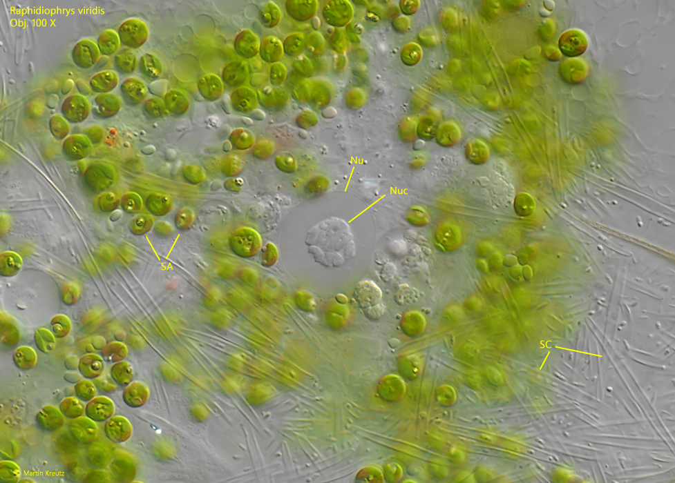
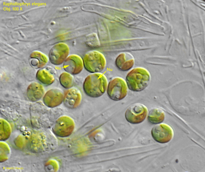
globular nucleus with central nucleolus
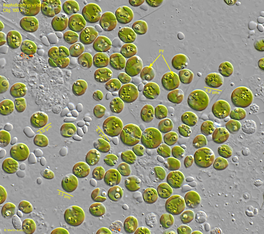
diameter symbiotic algae 5–6.5 µm
numerous axopodia, about length of colony
Raphidiophrys viridis
I find Raphidiophrys viridis among the floating plants in the Simmelried. However, the species occurs only sporadically. I have records from May 2005 and then again 20 years later in June-July 2025. I found the colonies particularly frequently in old samples and on the walls and bottom of the vessels.
The colonies of Raphidiophrys viridis are very conspicuous in the samples, with a diameter of about 200 µm and green-colored cells (s. figs. 1, 2 and 3). The cells lie close together in the colonies and do not form long, slender plasma bridges like Raphidiophrys elegans. However, the essential distinguishing feature are the scales covering the colonies, which in Raphidiophrys viridis are elongated rods with tapered ends. They resemble the shape of a boat (s. figs. 9 and 10). In my population, the scales were 25–35 µm long. They are said to have fine striations and small papillae at the poles, but I was unable to detect these.
According to my observations, the nucleus has a large, central nucleolus (s. fig. 6). The numerous symbiotic algae in the cytoplasm have a slightly irregular shape and a diameter of 5.0–6.5 µm. They have a pyrenoid and their own cell nucleus (s. figs. 7 and 8). In addition, the cytoplasm always contained some highly refractive granules. They do not appear to belong to the genus Chlorella. The symbiotic algae of Rhaphidiophys viridis resemble those of Raphidiophrys elegans.
Fig. 1:Raphidiophrys viridis. D = 230–240 µm (of colonies). The colonies between detritus particles. Obj. 20 X.
Fig. 2:Raphidiophrys viridis. D = 290 µm (of colony). A colony with fully extended axopodia. Obj. 20 X.
Fig. 3:Raphidiophrys viridis. D = 260 µm (of colony). A colony in brightfield illumination. Obj. 40 X.
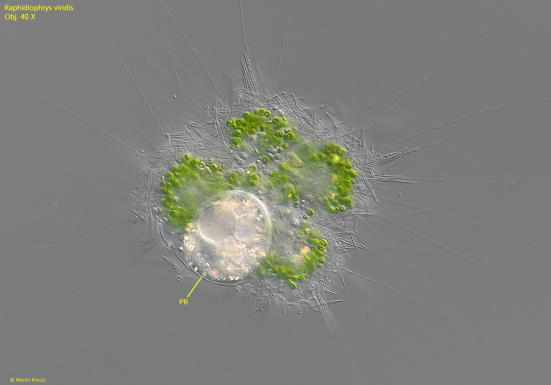
Fig. 4:Raphidiophrys viridis. A colony with catched prey (PR). The prey is a ciliate. Obj. 40 X.
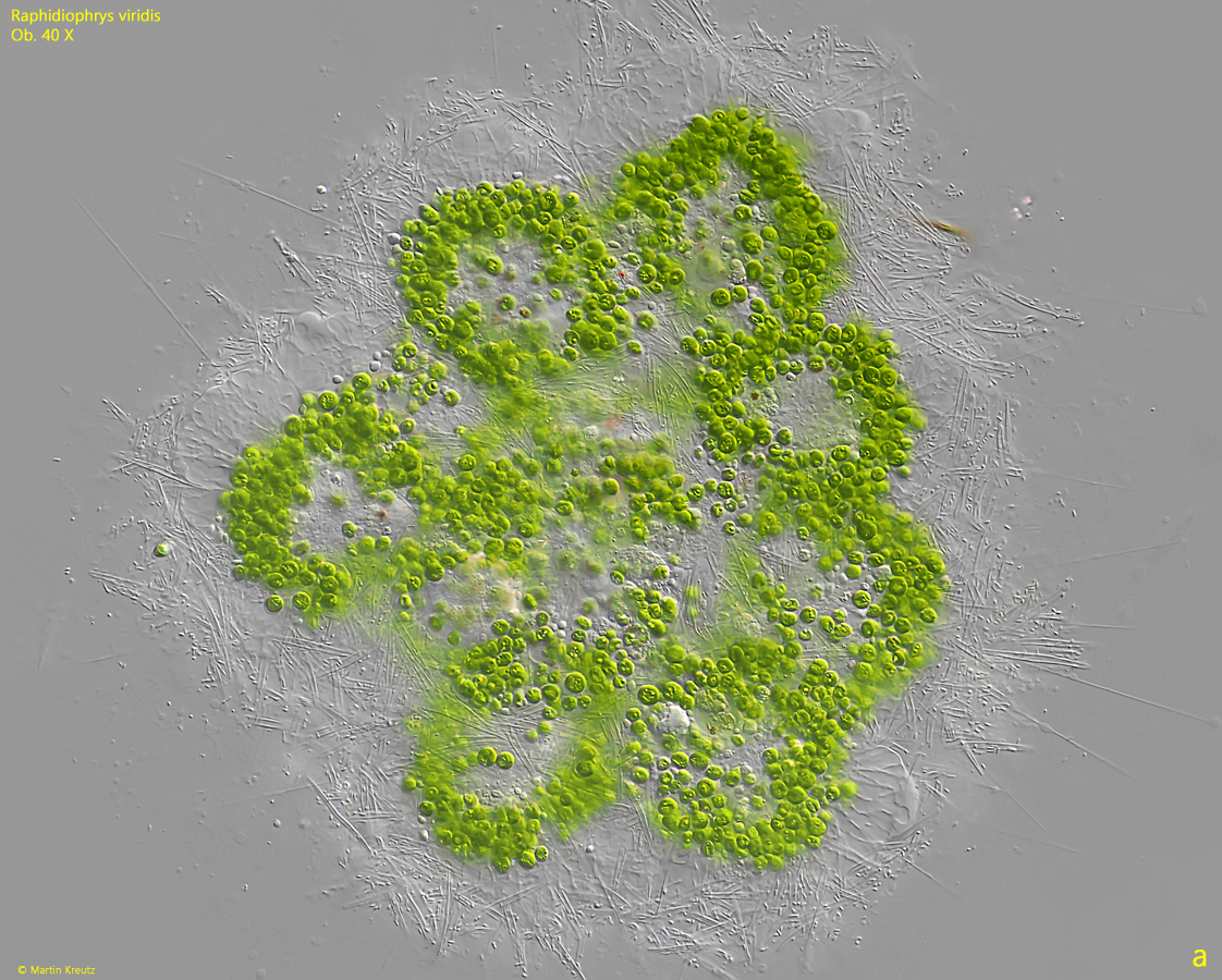
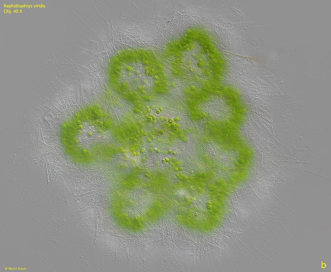
Fig. 5 a-b:Raphidiophrys viridis. Two focal planes of a slightly squashed colony. Obj. 40 X..
Fig. 6:Raphidiophrys viridis. The nucleus (Nu) with the central nucleolus (Nuc) in a squashed specimen. SA = symbiotic algae, SC = scales. Obj. 100 X.
Fig. 7:Raphidiophrys viridis. The symbiotic algae in a strongly squashed specimen. The symbiotic algae have a slightly irregular shape and a diameter of 5.0–6.5 µm. A pyrenoid (PY) is present. The algae are not of the Chlorella type. Obj. 100 X.
Fig. 8:Raphidiophrys viridis. The symbiotic algae of a second specimen with the visible nucleus (Nu). Obj. 100 X.
Fig. 9:Raphidiophrys viridis. The rod-shapes scales with tapered ends of a strongly squashed specimen. The scales are 29–34 µm long. Obj. 100 X.
Fig. 10:Raphidiophrys viridis. The scales of a second, strongly squashed specimen. Obj. 100 X.