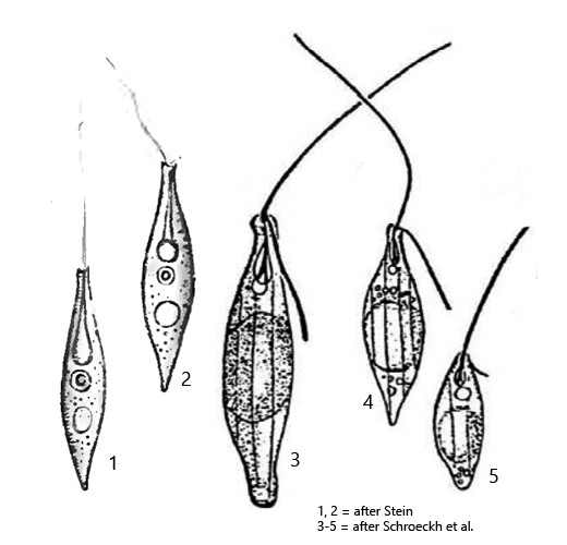
body elongate elliptical, posterior end pointed or slightly swollen
apically oblique with slight neck and collar
length 11–30 µm
pellicle with 6–8 inconspicuous, longitudinal ridges (hard to see)
locomotion flagellum with body length
trailing flagellum about one fourth of body length
reservoir dorsal with assoiciated contractile vacuole
nucleus above cell equator
gelatinous body in below nucleus
roundish paramylon grains below and above gelatinous body
Sphenomonas teres
My only find of Sphenomonas teres to date comes from the uppermost mud layer of the Simmelried. As this euglenid flagellate is quite small, I may have overlooked it in the past.
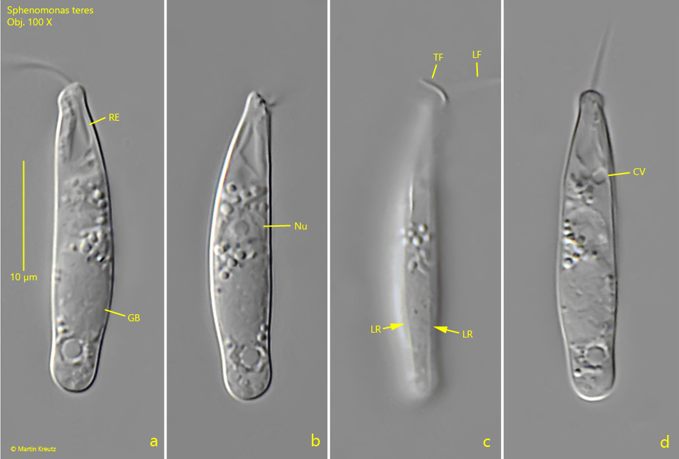
Sphenomonas teres can be distinguished from the similar species Sphenomonas quadrangularis by the fact that the pellicle has only a few delicate longitudinal ridges, which are difficult to recognize (s. fig. 3 c). In most cases the pellicular of Sphenomonas teres appears smooth. From the oblique apical end the locomototion flagellum and the trailing flagellum originate.
Almost all specimens in my population had a slightly swollen posterior end and thus resembled drawing 3 (s. drawings above). The members of the genus Sphenomonas have a large, gelatinous body, which is located approximately in the middle of the body (s. fig. 3 a). Sphenomonas teres also has such a body. This hyaline body has already aroused the interest of the first observers. It is insoluble in both alcohol and ether (i.e. no oil or fat). In water, sodium hydroxide and ammonia the body swells but does not dissolve. It is thought to be a high molecular weight reserve of unknown nature.
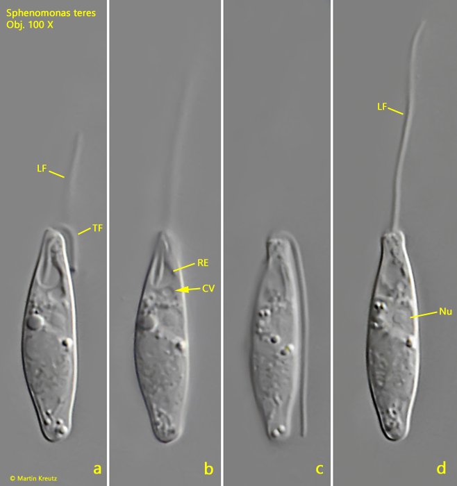
Fig. 1 a-d:Sphenomonas teres. L = 20 µm. A freely swimming specimen. Note the contractile vacuole (CV) associated with the reservoir (RE). LF = locomotion flagellum, Nu = nucleus, TF = trailing flagellum. Obj. 100 X.
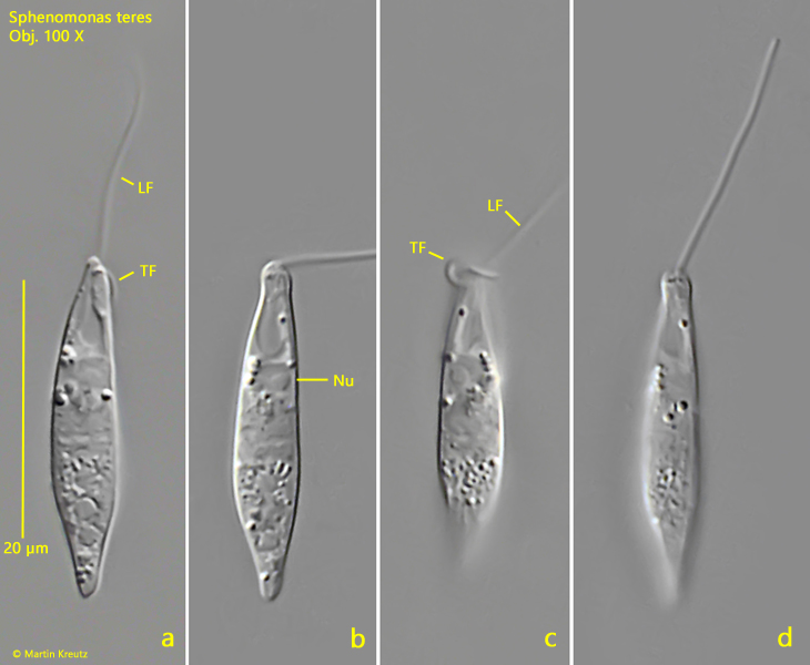
Fig. 2 a-d:Sphenomonas teres. L = 24 µm. A second, freely swimming specimen. LF = locomotion flagellum, Nu = nucleus, TF = trailing flagellum. Obj. 100 X.
Fig. 3 a-d:Sphenomonas teres. L = 27 µm. A third, freely swimming specimen from left (a), from right (b, c) and from dorsal (d). Note the delicate longitudinal ridges (LR) and the gelatinous body (GB) of unknown function. CV = contractile vacuole, LF = locomotion flagellum, Nu = nucleus, RE = reservoir, TF = trailing flagellum. Obj. 100 X.