body elongated trumpet-shaped, contracted obovoid to club-shaped
appears blue-green, no symbiotic algae
blue-green color due to granueles arranged in stripes beneath pellicle
length up to 4 mm (of elongated specimens)
adoral membranelle running in clockwise to oral funnel
attached with thigmotactic cilia to the substrate
macronucleus moniliform of about 6–20 spherical parts
12–42 micronuclei scattered near to the nodules of the macronucleus
contractile vacuole on left wall of oral funnel
Stentor coeruleus
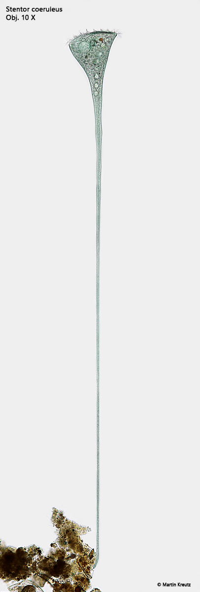
I find Stentor coeruleus frequently and regularly, especially in the Simmelried and Ulmisried. The specimens are easy to recognize due to their size and striking blue-green colouring. In fresh samples, the specimens usually swim around in a contracted form. In a micro-aquarium with a high layer thickness, however, they quickly begin to attach themselves to detritus particles and stretch out. Stentor coeruleus shows an amazing ability to stretch. When fully elongated, most of the body is almost tapered like a thread, except for the trumpet-shaped “head” (s. figs. 1 and 2). The elongated body bends back and forth as food is swirled towards it. Single-celled algae, dinoflagellates and bacteria serve as food.
The macronucleus is moniliform and consists of 10-20 spherical nodules (s. figs. 3 a and 5). This makes it easy to distinguish Stentor coeruleus from Stentor multiformis, which is also blue-green in color. The micronuclei of Stentor coeruleus are attached to the moniliform macronucleus, but also in the adjacent cytoplasm (s. fig. 6). According to my observations, the micronuclei are elliptical in shape and about 5–6 µm long (s. fig. 6).
Fig. 1:Stentor coeruleus. L = 1890 µm. A fully elongated specimen in brightfield illumination. Obj. 10 X.
Fig. 2:Stentor coeruleus. L = 1890 µm. The same specimen as shown in fig. 1 at higher magnification and with DIC. Obj. 20 X.
Fig. 3:Stentor coeruleus. Two focal planes of the trumpet-shaped anterior end. Note the adoral zone of membranelles (AZM) and the contractile vacuole (CV). Ma = nodules of the moniliform macronucleus. Obj. 40 X.
Fig. 4:Stentor coeruleus. Stripes of blue-green granules are arranged between the longitudinal rows of cilia. Obj. 100 X.
Fig. 5:Stentor coeruleus. The spherical nodules of the moniliform macronucleus (Ma) in a squashed specimen. Obj. 100 X.
Fig. 6:Stentor coeruleus. The small, elliptically shaped micronuclei (arrows) are scattered in the cytoplasm near the nodules of the macronucleus (Ma). Obj. 100 X.