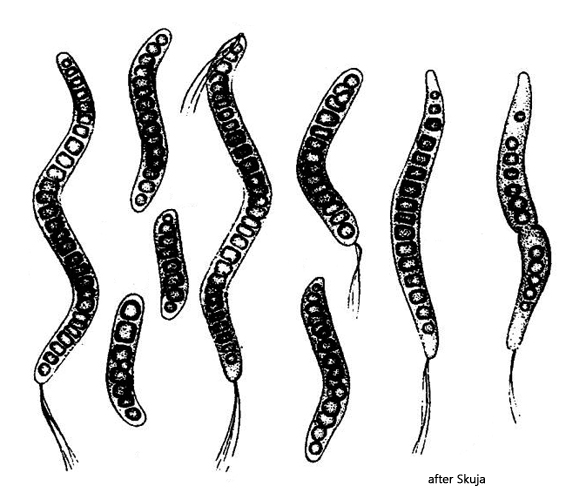
I found large numbers of Thiospira winogradskyi in samples from the Schwemm Moor (Austria). The only accurate description of this sulfur bacterium appears to be that provided by Skuja (1956).
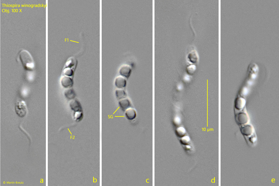
The species within the genus Thiospira are characterized by a spiral-shaped body and sulfur globules, which are always arranged in a row within the cells. In addition, the cells have flagella at one or both ends, which are often twisted into bundles.
The individual species within the genus Thiospira differ mainly in terms of cell diameter and length. The cells in my population had a diameter of 2.5–3.0 µm. This rules out the species Thiospira tenuis, which has a diameter of 0.8–1.0 µm, and Thiospira dextrogyra, which has a diameter of 1.0–1.3 µm. The species Thiospira bipunctata has two conspicuously large sulfur globules and no globules arranged in a row. This leaves the species Thiospira winogradskyi, which is said to have a diameter of 1.5–3.5 µm. This corresponds to the diameter of the cells in my population. However, Skuja specifies a cell length of 20–60 µm for Thiospira winogradskyi, while the cells in my population were 12-22 µm long. However, since there are no alternatives to Thiospira winogradskyi and the cells in my population correspond to Skuja’s drawings (s. above), I am sticking with this species.
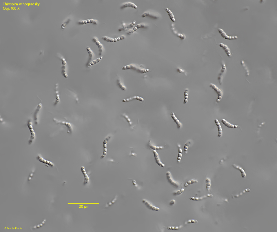
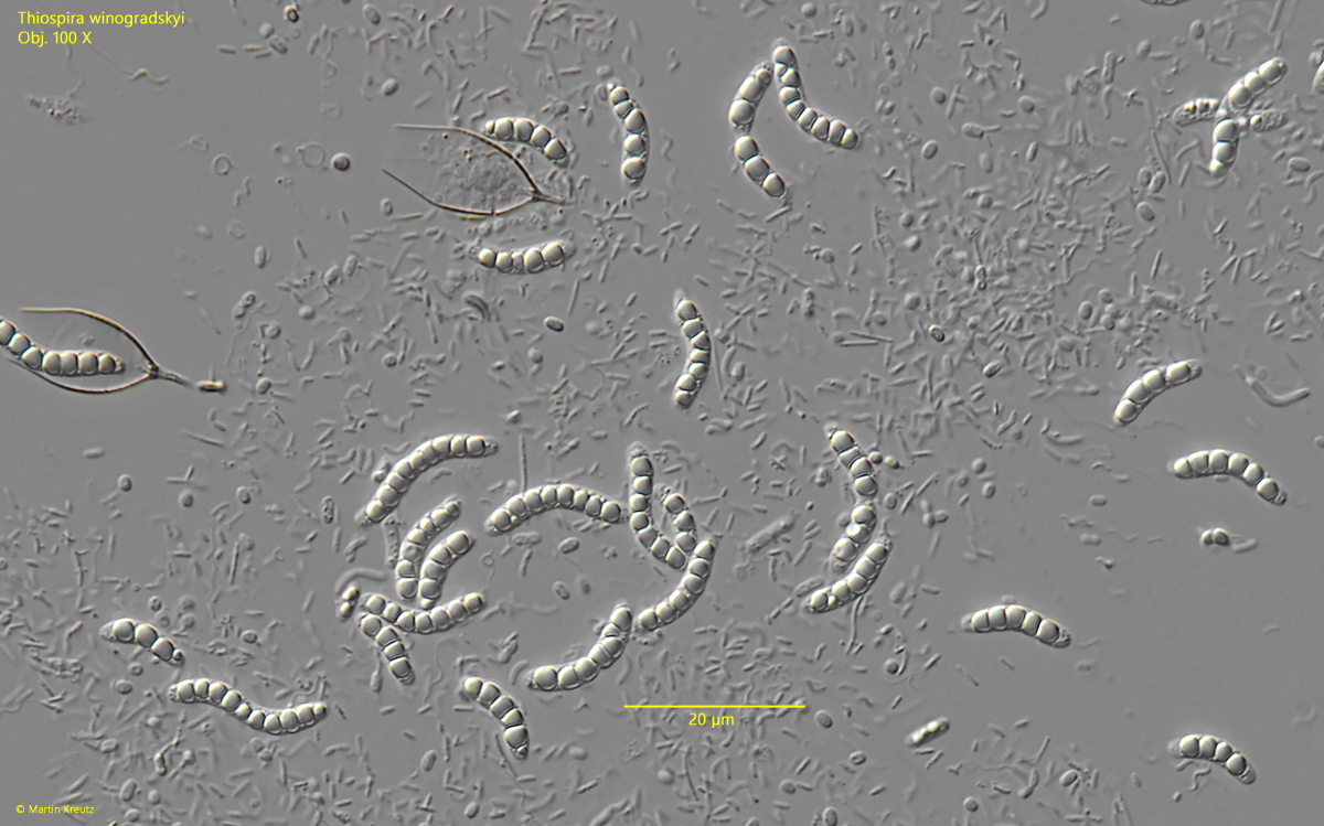
Fig. 1:Thiospira winogradskyi. L = 13–21 µm. An accumulation of freely swimming specimens. Obj. 100 X.
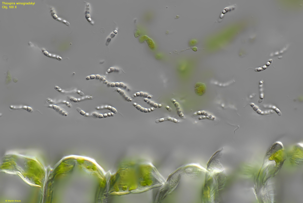
Fig. 2:Thiospira winogradskyi. L = 11–21 µm. A second accumulation of freely swimming specimens. Obj. 100 X.
Fig. 3:Thiospira winogradskyi. L = 12–19 µm. Some slightly squashed specimens. Obj. 100 X.
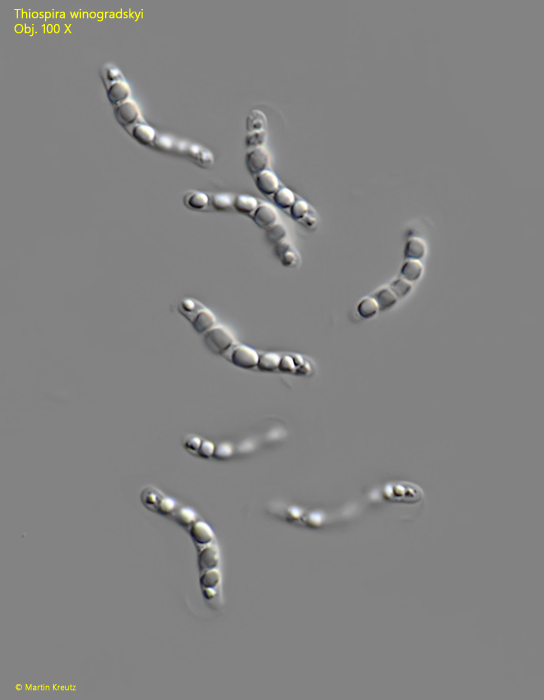
Fig. 4 a-e:Thiospira winogradskyi. L = 13–22 µm. Some specimens in detail. Each cell has a polar bundle of flagella at both ends (F1, F2). In the cells large sulphur globules (SG) are arranged in a row. Obj. 100 X.
Fig. 5:Thiospira winogradskyi. L = 13–18 µm. Some specimens scattered in a detritus flake. Obj. 100 X.