Tortitaenia obscura is described by Lenzenweger (2003) as widespread. I was only able to find Tortitaenia obscura once in June 2026 in the Schwemm bog in Austria. However, the species was extremely rare in the samples.
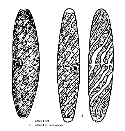
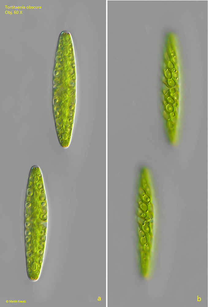
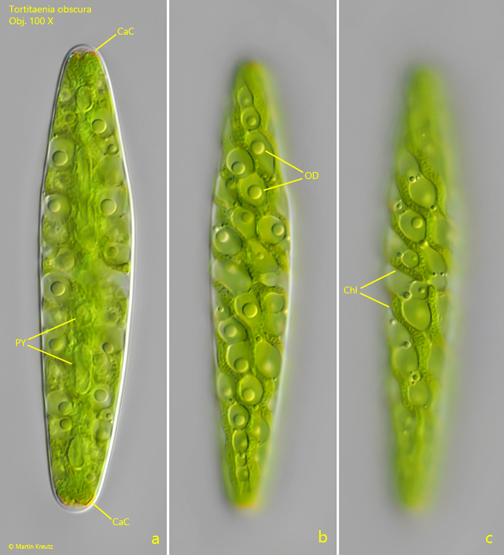
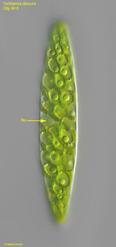
The cells of Tortitaenia obscura are usually found in pairs within a common mucus envelope (s. fig. 1 a-b). They are oriented longitudinally to each other and slightly offset. With an average length of about 100 µm, the cells are quite large. However, their size is very variable. The cell shape can be described as loaf-shaped. It is not spindle-shaped like the similar species Spirotaenia erythrocephala. A key feature of Tortitaenia obscura are the 3–8 ribbon-shaped chloroplasts, which are arranged spirally. Their margin is irregularly wavy. Numerous oil droplets are located between the chloroplasts (s. fig. 2 b). I found the nucleus laterally shifted from the longitudinal cell axis (s. fig. 3)
I found all the pyrenoids arranged along the longitudinal cell axis (s. fig. 2 a). Some of the pyrenoids were elongated in shape. This arrangement contradicts the description by Lenzenweger (2023), who observed the pyrenoids scattered among the chloroplasts. It is difficult to say whether he accidentally identified the oil droplets between the chloroplasts as pyrenoids. All other features match the description of Tortitaenia obscurum by Lenzenweger.