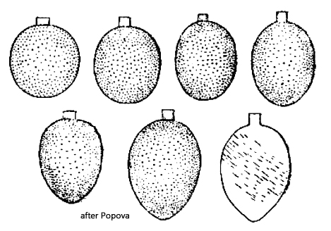
So far, I have found Trachelomonas manginii f. subpunctata exclusively in the Simmelried. The shape of the lorica shows some variability. I have mainly found specimens with an almost spherical lorica (s. fig. 3 a-c) and ellipsoid specimens (s. figs. 1 a-c and 2 a-c). All specimens had a comparatively narrow neck, which is clearly set off from the lorica and has a cylindrical or slightly conical shape. The apical margin of the neck is smooth, without spines. The lorica has a uniform thickness and all specimens I examined had distinct punctation, as Popova (1966) illustrated (s. drawings above).
My specimens were about 10% larger than those investigated by Popova but corresponded in all other characteristics to the description of Trachelomonas manginii f. subpunctata. The basic form Trachelomonas manginii has a smooth shell without punctation but is otherwise identical.
The similar species Trachelomonas planctonica has a strongly punctuated or scrobiculate (furrowed, pitted) thick-walled lorica with a spherical or broadly ellipsoid shape and a cylindrical neck. The neck is 4–5 µm wide and 2.6–4 µm high (Huber-Pestalozzi, 1955). Popova also reports a neck height of up to 4 µm. In my specimens, the neck was 3.6–4.2 µm wide and 2.4–2.9 µm high, thus narrower and shorter. It also had a smooth, non-serrated apical rim, and the thin-walled lorica had only fine punctation. These features indicate Trachelomonas manginii f. subpunctata.
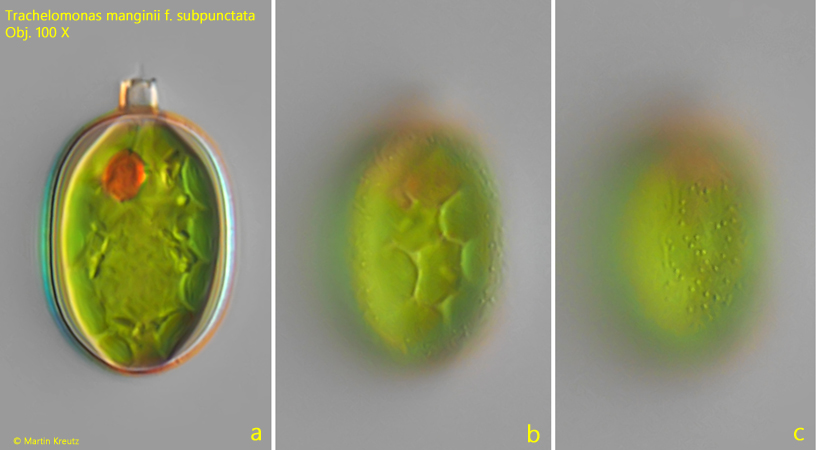
Fig. 1 a-c:Trachelomonas manginii f. subpunctata. L = 28 µm (with neck). Three focal planes of a freely swimming specimen. Note the dottet surface of the lorica (c). Obj. 100 X.
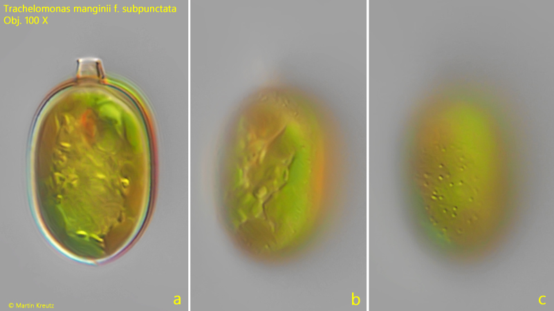
Fig. 2 a-c:Trachelomonas manginii f. subpunctata. L = 30 µm (with neck). A second, freely swimming specimen. Obj. 100 X.
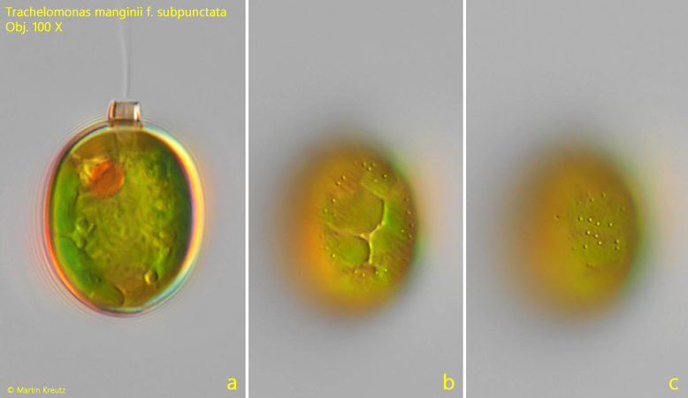
Fig. 3 a-c:Trachelomonas manginii f. subpunctata. L = 27 µm (with neck). Three focal planes of a third specimen. Obj. 100 X.