How I use immersion lenses

Visitors to my website “realmicrolife.com” may notice that around 70% of all images shown were photographed using my immersion lenses U-PlanNApo 60X/1.42 and U-PlanSApo 100X/1.4. I use these two lenses most frequently because they project objects as large as possible onto the camera sensor while also offering high resolution. Many identifying features of protozoa and metazoans can only be seen and displayed with immersion lenses.


Many microscopists have a certain aversion to using immersion lenses because both the condenser and the lens must be immersed with immersion oil. The oil on the condenser front lens is particularly critical because it can flow between the slide and the cross table when the slide holder is moved. Cleaning is then time-consuming.


That’s why I’ve been using water to immerse the condenser for years, namely normal tap water, and only immersion oil for the lens. For 30 years, I have always used the same immersion oil from Merck with the item number 1.04699.0100 and a specified refractive index of nD20 = 1.515-1.517 (s. fig. 1). For use, I always pour some into a 5 ml dropper bottle. This is fitted with a small collar made of filter paper (coffee filter) to prevent excess oil from running off (s. fig. 1).

Use immersion objectives

Fig. 1: The Merck immersion oil and the 5 ml dropper bottle I use to measure out the oil.

The use of water with a refractive index of nD20 = 1.33 for immersion may seem unusual to some readers, but the effect on resolution is not as great as one might think. Below, I explain this using the Abbe equation for microscope resolution.


In practice, I first place a drop of water on the condenser lens using a disposable PE pipette. I do this while the slide is already on the cross table (s. fig. 2 a). Then I place a small drop of immersion oil on the slide where the object is located. I normally do this with the immersion lens slightly swung out. In fig. 3 a-b, I have applied this in front of the lens to illustrate the volume of oil applied (s. fig. 3 b). I always use very little oil, about 10-30 µl. I only use more oil when I want to track moving objects. 

Use immersion objectives

Fig. 2 a-b: Immersion of the condensor frontlens with water. 

Use immersion objectives

Fig. 3 a-b: Application of the immersion oil on the coverslip. 

Use immersion objectives

Fig. 4 a-b: Final water/oil immersion.

After observation, I remove the water from the front lens of the condenser with a soft cotton cloth (s. fig. 5). I would like to mention here that even after well over 1000 immersions with water, I have not observed any negative effects on the condenser lens.

Use immersion objectives

Fig. 5: Removal of the water from the condensor front lens with a cotton cloth.

What about the resolution of the microscope when using water on the condenser side? This can be calculated using Abbe’s equation for microscope resolution (s. also Abbe equation):


d = 1.22 λ / (NAObj + NACond)


where


d = the minimum distance between two points that can be resolved
λ = wavelength of light, standard is green 550 nm (= 0.55 µm)
1.22 = factor that takes into account the dimension of the Airy discs

NAObj = aperture of the lens

NACond = aperture of the condenser


My condenser has NA = 1.4 and the 100 X lens also has NA = 1.4. With oil/oil immersion and a refractive index of nD20 = 1.516, I can use the full aperture of the condense and the lens. This results in:


doil/oil = 1.22 X 0.55 µm / (1.4 + 1.4)

doil/oil = 0.239 µm


If I use a water/oil immersion, the condenser can achieve the following maximum numerical aperture:


NACond = nD20 water X sin α


with


α = one-half opening angle


The half opening angle α is 67.7° for a condenser with NA = 1.4. This results in the following aperture when water is used as the immersion medium for the condenser:


NACond new = 1.33 X sin 67.7

NACond new = 1.23


This value can now be inserted into the Abbe equation:


dwater/oil= 1.22 X 0.55 µm (1.23 + 1.4)

dwater/oil = 0.255 µm


This results in a deterioration in resolution of 0.016 µm, which is only 6.7% less than with oil/oil immersion.


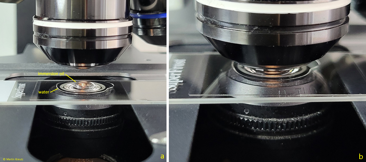
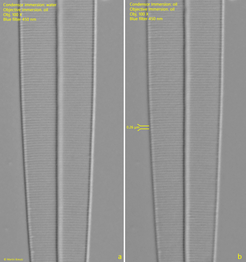
Is this deterioration of 6.7% relevant in practice? To find out, I first photographed the shell structure of Amphipleura pellucida with a line spacing of 0.26 µm using water/oil immersion (s. fig. 6 a) and then photographed the same area of the specimen using oil/oil immersion (s. fig. 6 b). In both cases, I used a 450 nm blue filter.

Use immersion objectives

Fig. 6 a-b: The resolution of the shell structure of Amphipleura pellucida with water/oil immersion (a) and oil/oil immersion (b). 

There is practically no difference. This means that for my typical applications, observing protozoa and metazoans in an aqueous medium, the 6.7% reduction in maximum possible resolution is completely insignificant. With water/oil immersion, I get flawless, high-resolution results. In addition, water/oil immersion is much more practical to implement and eliminates the need for time-consuming cleaning.