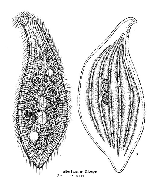
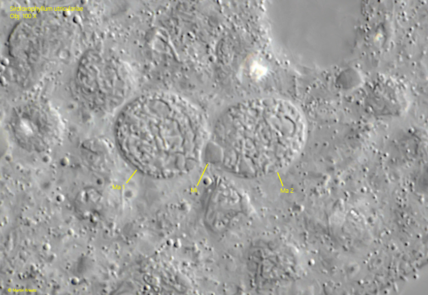
two macronuclear nodules and a spherical micronucleus between them
two contractile vacuoles in anterior and posterior third
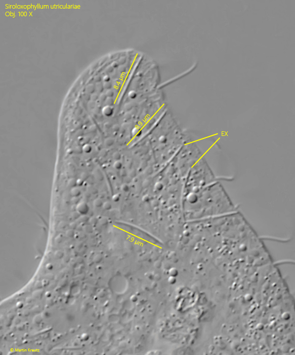
extrusomes 6–8 µm long, slightly curved
Siroloxophyllum utriculariae
Siroloxophyllum utriculariae was first described by Penard (1922) as Amphileptus utriculariae and was later transferred by Kahl (1926) to the genus Loxophyllum, because he saw the naked left side and the fringe of extrusomes, which runs along both the ventral and dorsal margins. Finally, in 1995 the species was transferred by Foissner & Leipe to Spiroloxophyllum on the basis of the shape of the dorsal brush and a winding structure of the ventral and dorsal margins.
I find Siroloxophyllum utriculariae only very rarely. In August 2003, in the Mühlweiher Litzelstetten, I found only a single specimen among floating plants. Only 23 years later, in September 2026, I found a large population of Siroloxophyllum utriculariae in a water-filled stone basin in the garden of the Inzighofen monastery. The photos shown below depict specimens from this population.
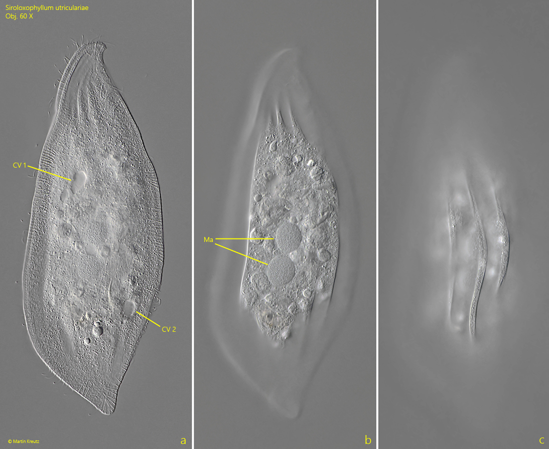
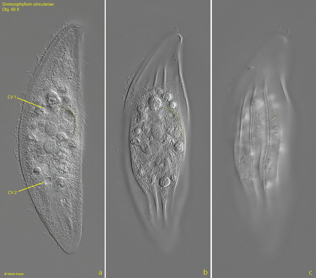
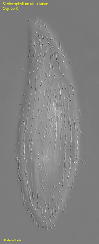
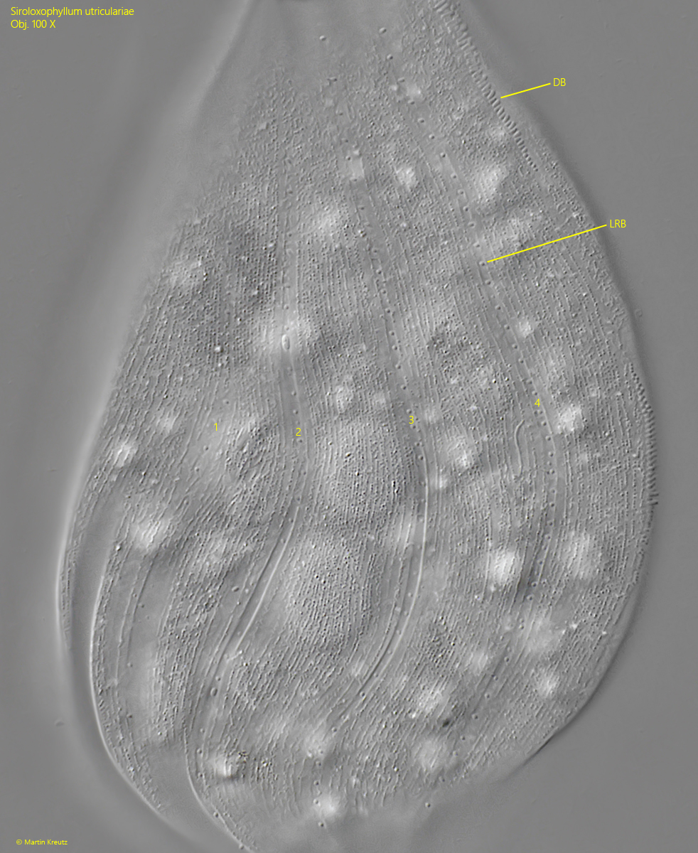
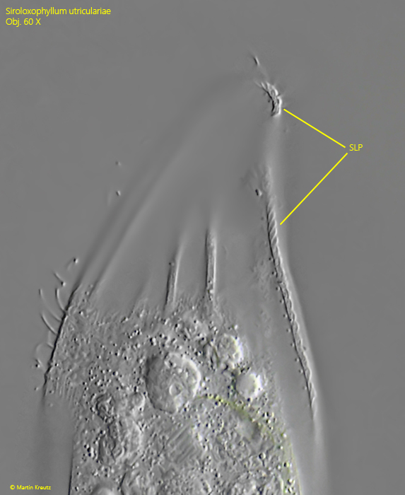
Siroloxophyllum utriculariae is strongly flattened laterally and leaf-shaped. The ciliate is also very hyaline. The right side is flat and has 14–15 rows of cilia. The left side is convex in the middle with 3–8 distinct ridges (s. fig. 1 c). Between the ridges there are 4 rows of short bristles (s. fig. 5). The entire margin of the ciliate has the twisted pattern of a rope, which is a characteristic feature of this genus (s. fig. 6). In the middle of the body there are two round macronuclei, with the micronucleus positioned between them (s. fig. 7). As a characteristic feature, Siroloxophyllum utriculariae has two contractile vacuoles, which lie in the middle section of the body and are usually slightly offset diagonally (s. figs. 1 a and 2 a). Since they empty alternately, sometimes only one contractile vacuole is visible.
Siroloxophyllum utriculatriae can be distinguished from Loxophyllum meleagris and Loxophyllum helus because these species possess only one contractile vacuole and because the dorsal margin is occupied by wart-like bundles of extrusomes. Siroloxophyllum utriculariae differs from Litonotus species by the fringe of extrusomes on the ventral and dorsal margin (in Litonotus only ventral) and by the number of contractile vacuoles (in Litonotus only one terminal).
Fig. 1 a-c: Siroloxophyllum utriculariae. L = 217 µm. A freely swimming specimen from left. Note the two diagonally offset contractile vacuoles (Cv 1, CV 2) and the two globular macronuclei (Ma) in the center. Obj. 60 X.
Fig. 2 a-c: Siroloxophyllum utriculariae. L = 182 µm. A second freely swimming specimen from left. CV 1, CV 2 = contractile vacuoles. Obj. 60 X.
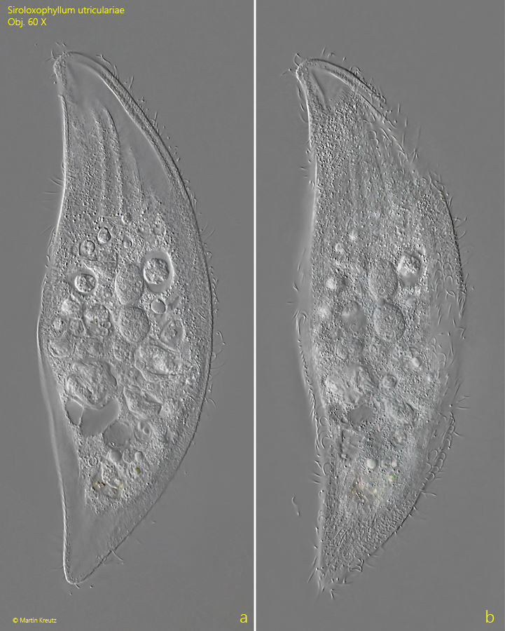
Fig. 3 a-b: Siroloxophyllum utriculariae. L = 230 µm. A third freely swimming specimen from right. Obj. 60 X.
Fig. 4: Siroloxophyllum utriculariae. L = 180 µm. Focal plane on the longitudinal rows on cilia of the right side. Obj. 40 X.
Fig. 5: Siroloxophyllum utriculariae. The left side of a squashed specimen. The flattened ribs show a fine longitudinal striation of granules. Between them lie 4 longitudinal rows of short bristles (1–4, LRB). DB = dorsal brush. Obj. 100 X.
Fig. 6: Siroloxophyllum utriculariae. The string-like pattern (SLP) of the oral bulge. Obj. 60 X.
Fig. 7: Siroloxophyllum utriculariae. The two globular macronuclei (Ma 1, Ma 2) with the micronucleus (Mi) in between. Obj. 100 X.
Fig. 8: Siroloxophyllum utriculariae. The extrusomes (EX) are slightly curved with a length of 7–9 µm.