cell spherical, diameter 24–65 µm (including layer auf scales)
covered with flattened, oval scales
scales oval in frontal view, length 3.0–5.4 µm X width 1.6–3.5 µm
scales slender oval in lateral view, in the middle often swollen.
cytoplasm orange or reddish with colored granules
feeds on algae
nucleus located eccentrically, with central nucleolus
no contractile vacuole visible
pseudopodia fine and short
No drawings from previous authors available.
I have found Pompholyxophrys stellata so far exclusively in the Simmelried. The findings are from January 1997, February 2012 and July 2014. After that I have not found any more specimens. The species can be easily distinguished from Pompholyxophrys puniceaby the flattened scales. In lateral view the scales appear very flat oval, sometimes somewhat thickened in the middle. In frontal view they are oval. Leaving these differences aside, the species appears to have the same characteristics as Pompholyxophrys punicea.
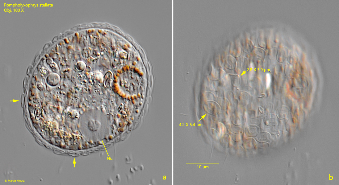
Fig. 1 a-b:Pompholyxophrys stellata. D = 40 µm (of the squashed specimen). Two focal planes of a slightly squashed specimen. Note that some of the flat scales in lateral view are “swollen” in the middle (a, arrwos). In frontal view the scales appear oval (b). Nu = nucleus. Obj. 100 X.
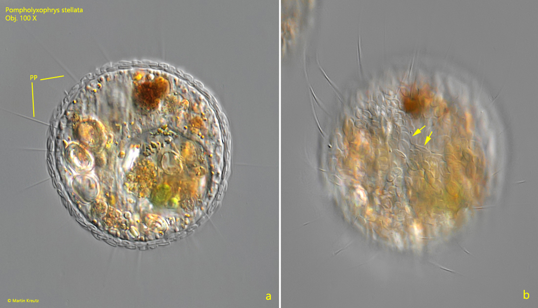
Fig. 2 a-b:Pompholyxophrys stellata. D = 54 µm (of the squashed specimen). Two focal planes of a slightly squashed second. In frontal view the flat scales are oval (b, arrows). PP = pseudopodia. Obj. 100 X.