Rhinothrix 1

Most likely ID: Rhinothrix nov. spec.


Synonym: n.a.


Sampling location: Ulmisried, Simmelried


Phylogenetic tree: n.a.


Diagnosis: 

  • body clavate, tapered to posterior end
  • length about 160–170 µm
  • oral bulge oblique with dorsal beak-shaped extension
  • ridge of extrusomes runs from oral bulge to right side of body
  • ridge of extrusomes ends near posterior third of right side
  • extrusomes type 1 curved rods, 14–16 µm long
  • extrusomes type 2, straight rods, 3.0–3.2 µm long
  • dorsal brush of three row, club-shaped cilia
  • macronucleus elongated ellipsoid
  • one spherical micronucleus
  • contractile vacuole terminal

No drawings from previous authors available.

So far, I have found three specimens of Rhinothrix 1. The first 2 specimens I found in the upper mud layer in Simmelried in May 2022. Only a few months later, I found a third specimen in October 2022 in Ulmisried. There, the specimen was also found in a sample of the bottom mud.


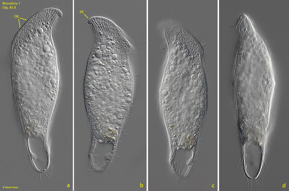
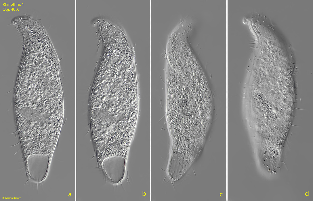
According to the definition by Foissner, Xu & Kreutz (2005), the genus Rhinothrix is characterized by a ciliation similar to the genus Aperthospathula and an oral bulge that has an ventral extensionen with a spiral course up to or almost to the posterior end. On the dorsal side the oral bulge has a nose- or finger-shaped extension. These features seem to be fulfilled in the spathidiid ciliate described here. However, its characteristics differ significantly from the three species described so far, Rhinothrix porculus, Rhinothrix barbatula, and Rhinothrix antennae, which is why it could be a previously undescribed species, which I would like to tentatively call Rhinothrix 1.


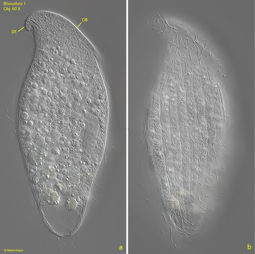
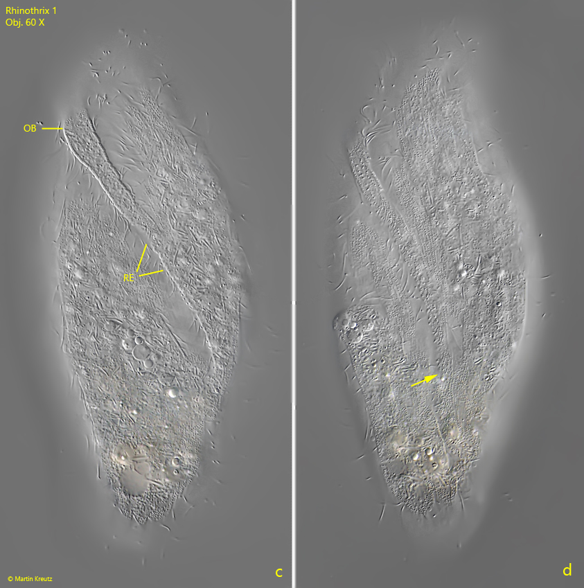
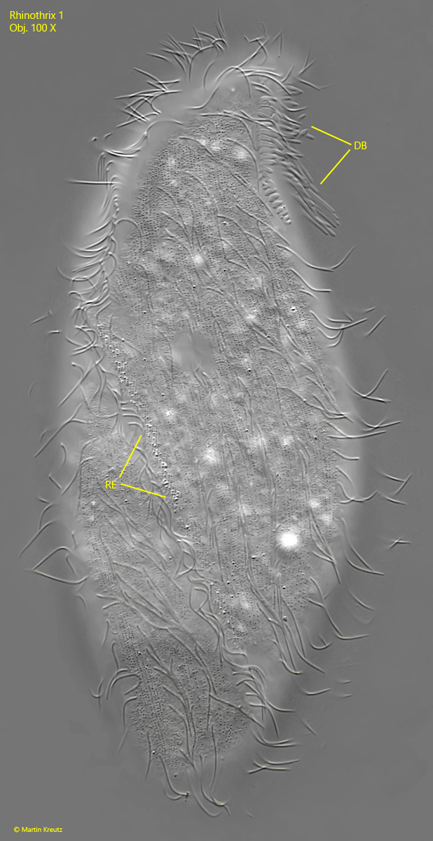
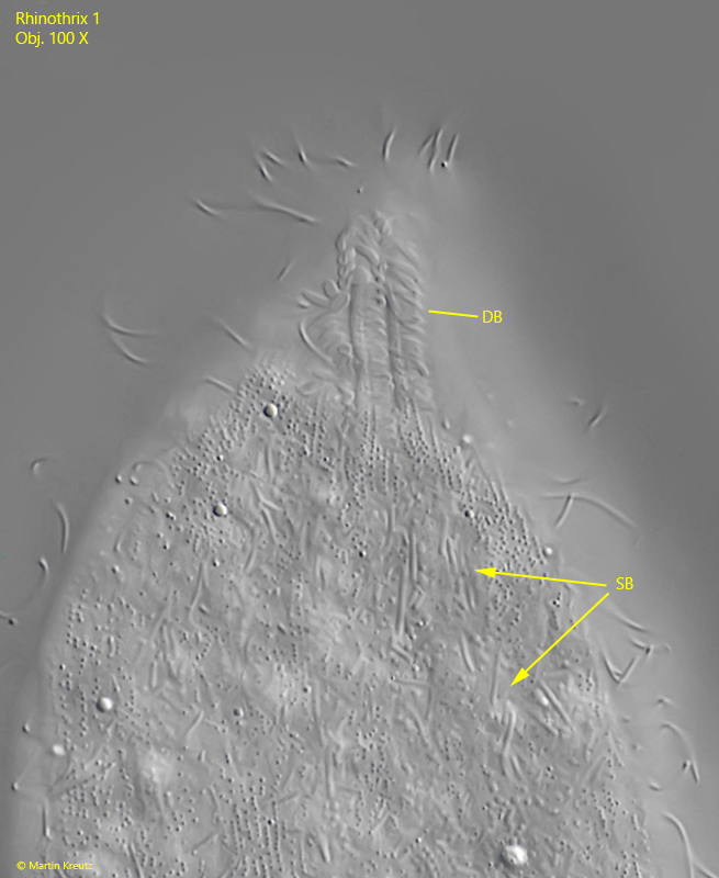
The specimens of Rhinothrix 1 had a length between 160-172 µm. The oral bulge is slanting ventrally and had a distinct, nose-shaped extension on the dorsal side (s. figs. 1 b and 3 a). Towards the ventral side, the oral bulge bends to the left side and flattens into a flat ridget that ends approximately in the posterior third of the body (s. figs. 3 c and 3 d). The ridge is accompanied on its left side by a parallel row of cilia (s. fig. 4).


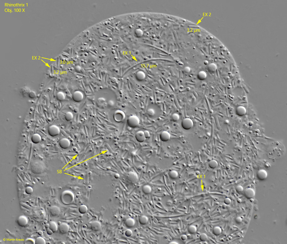
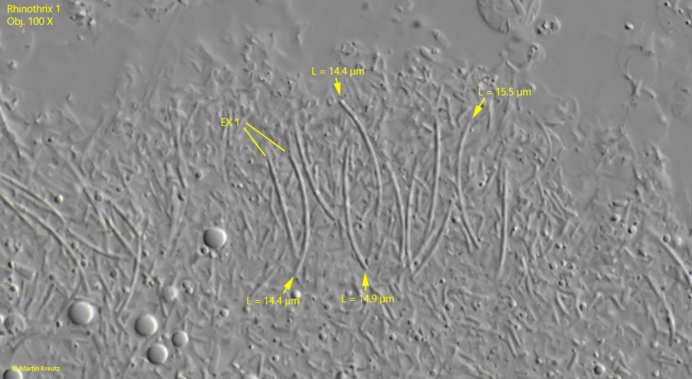
The oral bulge of Rhinothrix 1 is equipped with two types of extrusomes. The larger extrusomes of type 1 are distinctly curved and 14–16 µm long, while the extrusomes of type 2 are short, straight rods, with a length of 3.0–3.2 µm (s. figs. 8 and 9).


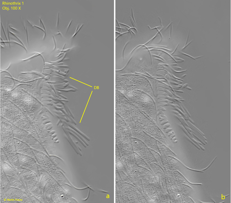
The dorsal brush of Rhinothix 1 consists of three rows, which are separated from each other by low ridges (s. figs 6 a-b and 7). The left row of the brush has the shortest cilia, which are elliptical or club-shaped. The right row has cilia up to 13 µm long, which are only slightly thickened at the distal end.


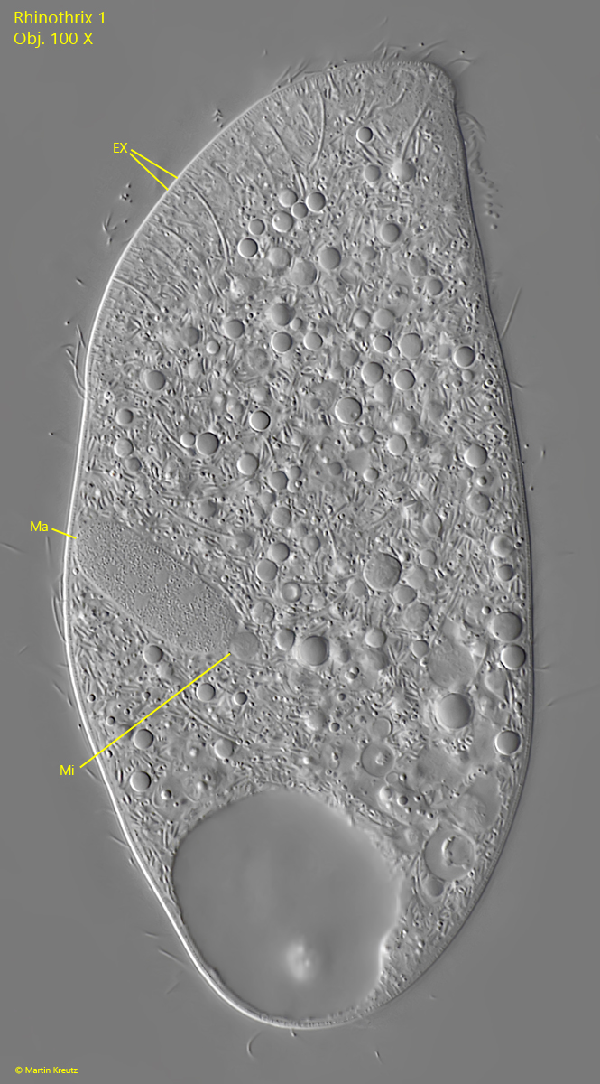
The macronucleus is located in the posterior half and is elongated ellipsoid. Attached to it is the spherical micronucleus. The contractile vacuole is terminal (s. fig. 5).

Rhinothrix-1

Fig. 1 a-d: Rhinothrix 1. L = 160 µm. A freely swimming specimen found in May 2022 in the Simmelried from left (a), right (b, c) and from ventral (d). Note the dorsal nose-shaped extension (DE) of the oral bulge (OB). Obj. 40 X.

Rhinothrix-1

Fig. 2 a-d: Rhinothrix 1. L = 172 µm. A second freely swimming specimen found in October 2022 in the Ulmisried from right (a, b, d) and from left (c). Obj. 40 X.

Rhinothrix-1
Rhinothrix-1

Fig. 3 a-d: Rhinothrix 1. L = 160 µm. A slightly squashed specimen as shown in fig. 1 a-d from right (a, b) and from left (c, d). The oral bulge extends ventrally into a ridge with extrusomes (RE), which runs to the left side and ends there in the last third of the body (arrow, d). The right side only shows the longitudinal rows of cilia (b). DE = dorsal nose-shaped extention of the oral bulge. Obj. 60 X.

Rhinothrix-1

Fig. 4: Rhinothrix 1. L = 160 µm. A slightly squashed specimen from left. The ridge of extrusomes reaches up to the posterior third of the body. Parallel to the ridge of extrusomes runs a row of cilia on its left side. DB = dorsal brush. Obj. 100 X.

Rhinothrix-1

Fig. 5: Rhinothrix 1. In a squashed specimen the elongated ellipsoid macronucleus (Ma) is visible as well as the adjacent spherical micronucleus (Mi). The oral bulge is equipped with two types of extrusomes (EX). Obj. 100 X.

Rhinothrix-1

Fig. 6 a-b: Rhinothrix 1. Two slightly different focal planes of the dorsal brush (DB). The longest cilia of the dorsal brush have a length of 13 µm. The shorter cilia are ellipsoid or club-shaped. Obj. 100 X.

Rhinothrix-1

Fig. 7: Rhinothrix 1. L = 160 µm. The dorsal brush (DB) in a view from dorsal. The three rows of the brush are separated by two flat ridges. SB = symbiotic bacteria scattered in the cytoplasm. Obj. 100 X.

Rhinothrix-1

Fig. 8: Rhinothrix 1. The extrusomes type 1 (EX 1) and type 2 (EX 2) in a strongly squashed specimen. The extrusomes of type 1 are curved and have a length of 14–16 µm while the extrusomes of type 2 are short, straight rods with a length of 3.0–3.2 µm. SB = symbiotic bacteria. Obj. 100 X.

Rhinothrix-1

Fig. 9: Rhinothrix 1. The curved extrusomes type 1 (EX 1) in a second squashed specimen. Obj. 100 X.